
|
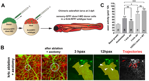
Fig. 7
(A) Diagram of the procedure for creating chimeric larvae to test where Duox1 functions to promote axon regeneration: cells from a duox1-MO injected sensory:GFP transgenic donor were transplanted into a wildtype Krt4:RFP transgenic host. GFP-labeled Rohon-Beard sensory neurons were thus deficient in Duox1 function, while RFP-labeled keratinocytes were wildtype. (B) Ablation of e3 wildtype keratinocytes (circle) and axotomy of a nearby duox1-morphant RB axon in the upper trunk region (arrow) promoted regeneration of severed axon branches (arrowheads) and reinnervation of denervated territory (shaded area) (see also Video S9). (C) Quantification of axon activity in chimeric embryos after keratinocyte ablation and axotomy, compared to basal growth (same as in Figure 1E), axotomy alone (same as in Figure 3D), and keratinocyte ablation (same as in Figure 4E). Sample size for each group is indicated by the numbers in the bars. Error bars represent the standard error of the mean. For statistical analyses, we performed one-way ANOVA and either Dunnett′s post-test to compare individual groups to controls (asterisks above bars indicate significance compared to control, the first column in graph) or Bonferroni′s post-test to compare individual groups with each other (as indicated by brackets, p = ns>0.05, * p<0.05, *** p<0.001).