
|
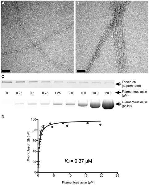
Fig. 2
Physical characterization of the interaction between fascin 2b and filamentous actin.
Negative staining and electron microscopy reveal actin bundling by recombinant fascin 2b fusion protein. Micrographs display filamentous actin in the absence of MBP-fascin 2b (A) and in the presence of MBP-fascin 2b (B); bundled filaments are observed in B. Scale bars are 100 nm. An immunoblot (C, top), from a cosedimentation experiment, shows the progressive depletion of MBP-fascin 2b from supernatants incubated with increasing concentrations of actin; molar concentration (C, middle) of actin for each lane is displayed. Image of a gel (C, bottom), after SDS-PAGE and exposure to SYPRO Ruby protein gel stain, shows the actin found in pellets from a cosedimentation experiment. Plotted are the means of specific equilibrium-binding measurements of MBP-fascin 2b to filamentous actin (D). Each point is a mean ± SEM (number of experiments, N = 4) or an average of two experiments (no error bars).