
|
Fig. 1
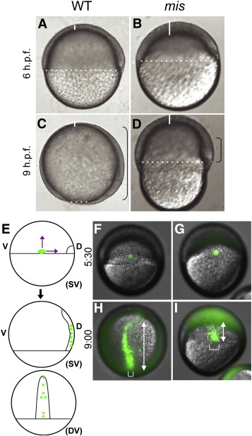
Gastrulation defects in maternally mutant mis embryos. Wild-type embryos (A,C) and mis mutant embryos (B,D). mis embryos exhibit reduced epiboly of the blastoderm, as evidenced by the increased thickness of the blastoderm at the animal pole (white solid lines) and reduced coverage of the yolk (white dotted line indicates blastoderm margin). The extent of involution of the hypoblast is also reduced (black brackets). Lateral views (animal top, dorsal right). (E–I) Defects in epiboly and convergence. (E) Labeling strategy, by uncaging a small group of cells at the lateral margin in wild-type (F) and mutant (G) embryos (SV: side view; DV: dorsal view). At a time corresponding to 90% epiboly in the wild-type, the extension of labeled cells along the animal–vegetal axis (double-headed arrows) is reduced in the mutant (I) compared to wild-type (H). In addition, the width of the band of labeled cells (brackets), indicative of cell intercalation during convergence, is reduced in mutant embryos. (F,G) are side views, dorsal to the right. (H,I) are centered on the labeled cells, corresponding to the dorsal view (DV) in (E). Uncaging was carried out at 5:30 h.p.f. and time after fertilization is indicated in h:min. Embryos shown in (F–I) are representative of 5 wild-type and 5 mutant embryos.
Reprinted from Developmental Biology, 353(2), Putiri, E., and Pelegri, F., The zebrafish maternal-effect gene mission impossible encodes the DEAH-box helicase Dhx16 and is essential for the expression of downstream endodermal genes, 275-289, Copyright (2011) with permission from Elsevier. Full text @ Dev. Biol.