
Image
|
Figure Caption
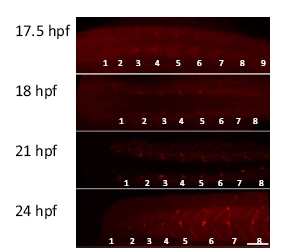
Fig. S3
Development of neuromuscular junctions. Fluorescently labeled α-bungarotoxin was used to label neuromuscular junctions during 17.5 to 24 hours postfertilization (hpf). Dorsal views (17.5 to 21 hpf) and a lateral view (24 hpf) are shown. Myotomes are numbered from rostral to caudal (1–9). (Scale bar, 50 μm.)
Acknowledgments
This image is the copyrighted work of the attributed author or publisher, and
ZFIN has permission only to display this image to its users.
Additional permissions should be obtained from the applicable author or publisher of the image.
Full text @ Proc. Natl. Acad. Sci. USA