
Image
|
Figure Caption
Fig. 7
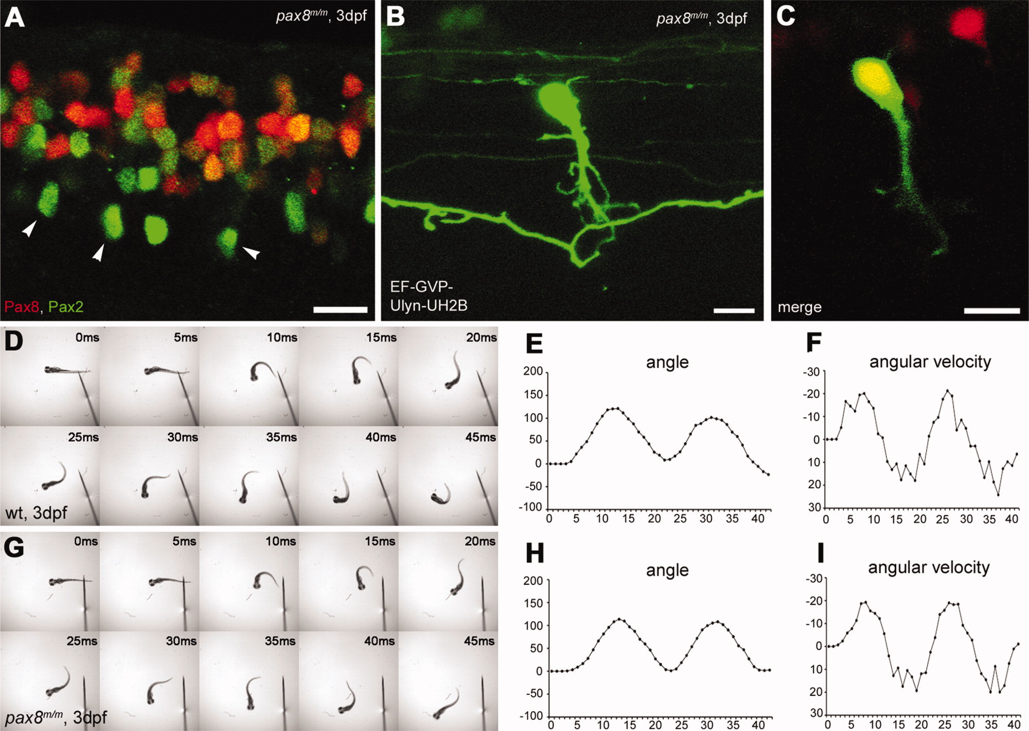
Spinal neurons in pax8 mutant. A: Spinal cord of a pax8m/m fish stained with anti-Pax2; RFP (red), anti-Pax2 (green). B,C: CoBL cell in pax8m/m fish. B is a stack of confocal images, and C is a single confocal plane; GFP (green), RFP (red). D–I: Touch response of wild-type (top, D–F) and pax8m/m fish (bottom, G–I) at 3 dpf. High-speed images of escape response are shown from 0 to 45 msec (D,G). In E,F,H,I, the head angle (E,H) and the angular velocity (F,I) were plotted against time. Magenta-green images of A–C are provided in Supporting Information Figure 5. Scale bars = 10 μm.
Acknowledgments
This image is the copyrighted work of the attributed author or publisher, and
ZFIN has permission only to display this image to its users.
Additional permissions should be obtained from the applicable author or publisher of the image.
Full text @ J. Comp. Neurol.