
Image
|
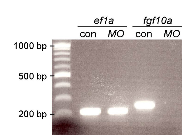
Figure Caption
Fig. S3
Validation of the fgf10a splice MO. Embryos were injected with 4 ng of the fgf10a MO, which targets the boundary of the 1st exon and 1st intron, harvested at 28 hpf, and used for RT-PCR analysis. fgf10a cDNA including the 2nd exon and part of the 1st and 3rd exons was amplified from control but not fgf10a MO-injected embryos, indicating that the MO blocks the proper splicing of fgf10a transcript. Elongation factor 1a cDNA was amplified from both control and fgf10a MO-injected embryos.
Acknowledgments
This image is the copyrighted work of the attributed author or publisher, and
ZFIN has permission only to display this image to its users.
Additional permissions should be obtained from the applicable author or publisher of the image.
Full text @ Development