
|
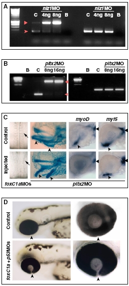
Fig. s12
Controls for the activity of MOs against POM genes. (A) RT-PCR on RNA from 30-hpf controls (C) or embryos injected with 4 ng (M4) or 8 ng (M8) of nlz1 MO, showing disrupted splicing of nlz1 mRNA (left lanes). Wild-type RNA is nearly absent in nlz1 morphants and replaced by immature mRNA retaining the first intron, which is predicted to contain an in-frame stop codon after 39 bp. The arrowhead and triangle point to the spliced and nonspliced nlz1 PCR products, respectively. EF1α (right lanes) was used as a loading control. B, blank reactions. (B) RT-PCR on RNA from 30-hpf controls (C) or embryos injected with 8 ng (M8) or 16 ng (M16) of pitx2 MO, showing disrupted splicing of pitx2 mRNA (left lanes). Wild-type RNA was nearly absent in pitx2 morphants and replaced by immature mRNA retaining the second intron, which is predicted to contain an in-frame stop codon after 30 bp, leading to a truncated protein lacking the last 14 aa in the homeodomain and the whole C-terminal region. The arrowhead and triangle point to the spliced and nonspliced pitx2 PCR products, respectively. EF1α (right lanes) was used as a loading control. B, blank reactions. (C Left) Disrupted somitogenesis in a 18s embryos injected with foxC1a MOs. (Center) Abnormal visceral arch morphogenesis in a 4-d-old embryo injected with pitx2 MO, as shown by cartilage staining (arrowheads point to derivatives of the first two arches). (Right) Eyes of 32-hpf hybridized controls or pitx2 MO-injected embryos showing reduction of myoD-positive ventral extraocular muscles (arrowheads), but not dorsal myoD-positive muscles (triangles) or myf5-positive muscles, in the pitx2 morphants. (D) Coloboma in foxC1A morphants is not caused by apoptosis. Lateral views (Left) of the head region of 72-hpf control embryos or embryos that were injected with a combination of foxC1a MOs and p53 MO. The choroid fissure (arrowheads) is not closed in embryos coinjected with foxC1a MOs and p53 MO. (Right) Images are higher magnifications of the eyes.