
|
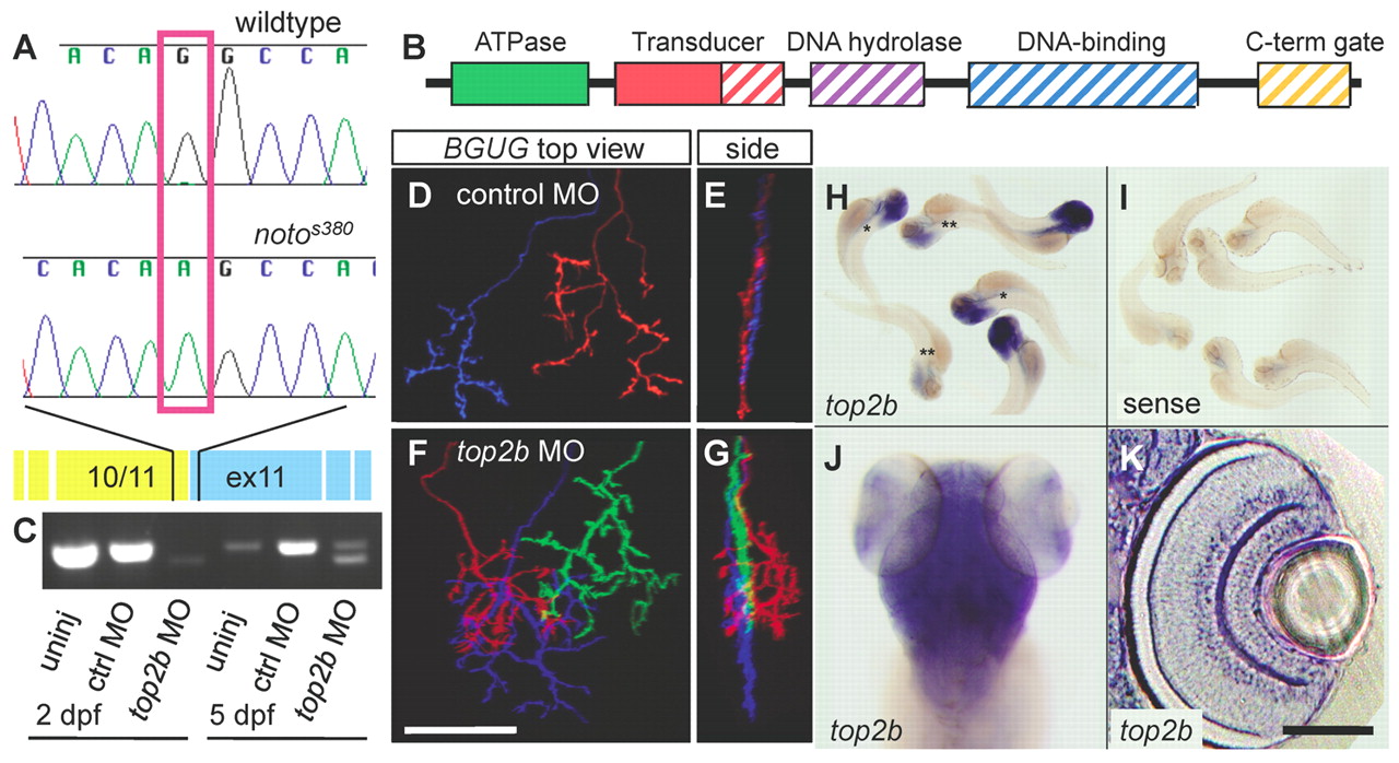
Fig. 4
The noto genetic lesion is a nonsense splice site mutation in top2b. (A) Sequence chromatograms showing the top2b exon 11 splice acceptor site in wild-type and mutant zebrafish. The sequence shown is at the intron-exon border shown in yellow and blue, respectively. (B) Schematic of the functional domains of Top2b protein. Solid colored regions are preserved in the predicted mutant protein; diagonal lines indicate the truncated portion. (C) RT-PCR-amplified top2b fragments from uninjected, control morphant and top2b morphant fish at 2 and 5 dpf. Morpholino oligonucleotide (MO) knockdown results in a truncated cDNA; both the full-length and truncated products can be detected by 5 dpf. (D-G) Ganglion cell (GC) axons from BGUG+ larvae in morpholino phenocopy experiment. (D,E) Top (D) and side (E) views of two SFGS axons from a control morphant. (F,G) Top (F) and side (G) views of three SFGS axons from an antisense top2b morphant; red axon is multilaminar. Scale bar: 50 μm. (H-K) In situ hybridization showing expression of top2b in 4 dpf larvae. (H) Antisense probe labels brain and eye. Unlabeled larvae are homozygous wild type (+/+), single asterisk indicates noto+/-, double asterisk indicates noto-/-.(I) Sense probe labeling is minimal. (J) Magnified dorsal view of antisense labeling. (K) Higher magnification view of sectioned retina, showing strong expression in amacrine cells (ACs) and GCs. Scale bar: 250 μm for H,I; 150 μm for J; 50 μm for K.