
Image
|
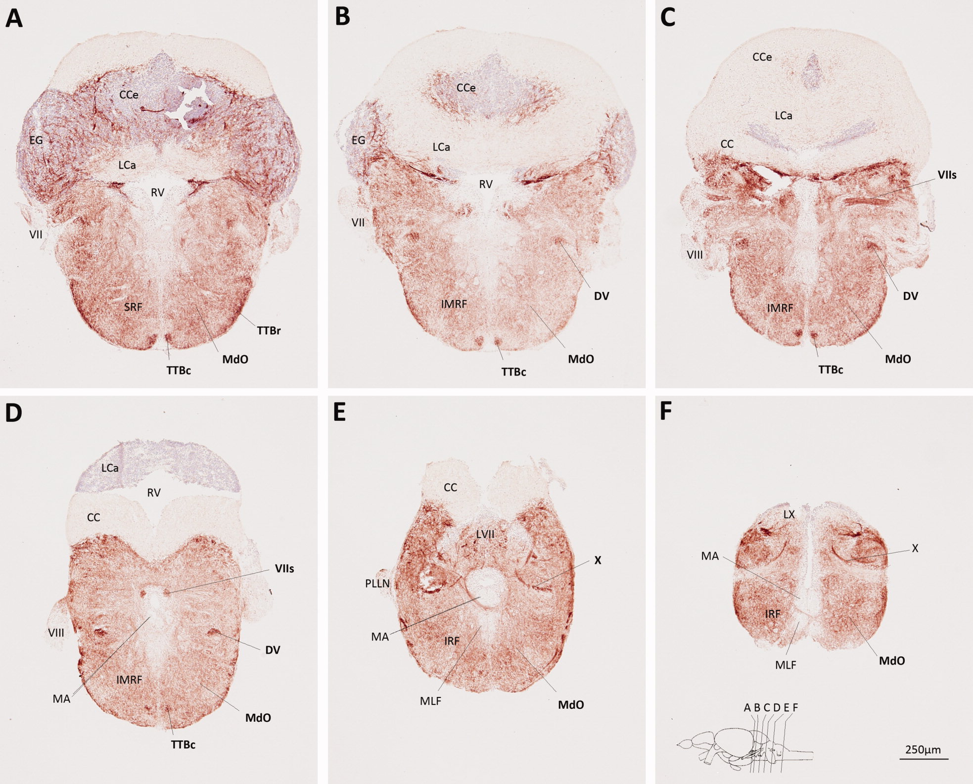
Figure Caption
Fig. 5
Myelin P0 expression in the hindbrain. Transverse sections of adult zebrafish hindbrain were labeled using the same method employed to generate Figure 3. Sections are oriented dorsal upwards and progress in a rostrocaudal direction; their planes are indicated in the inset to F, which also shows the scale bar for all six panels. Anatomical landmarks are indicated on the left side of each image and P0-immunoreactive structures are labeled in bold on the right side of each image.
Figure Data
Acknowledgments
This image is the copyrighted work of the attributed author or publisher, and
ZFIN has permission only to display this image to its users.
Additional permissions should be obtained from the applicable author or publisher of the image.
Full text @ J. Comp. Neurol.