
|
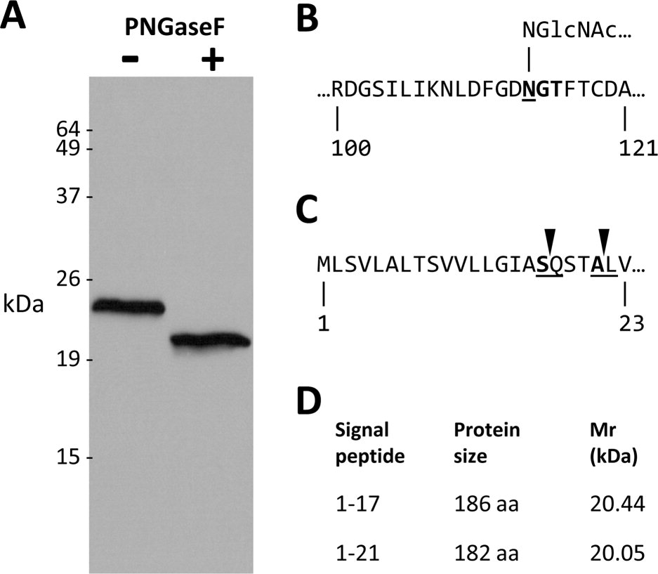
Fig. 2
Zebrafish P0 is a 23.5 kDa glycoprotein. A: A western blot probed with P0 antibody. Whole adult zebrafish brain lysate (32 μg protein per lane) was incubated with (+; right lane) or without (+ left lane) PNGaseF, prior to SDS-PAGE and western transfer. B: A short segment of the predicted extracellular domain of P0 is shown is shown (amino acids are numbered with respect to the full open reading frame). The consensus N-glycosylation motif is shown in bold. Underlining denotes the asparagine residue to which the N-acetyl-glucosamine side chain is linked. C: The N terminus of P0 is shown, illustrating the predicted signal peptide cleavage sites. D: The table shows the size and predicted molecular mass of P0 after cleavage of the signal peptide at each of the putative cleavage sites.