
|
Fig. 1
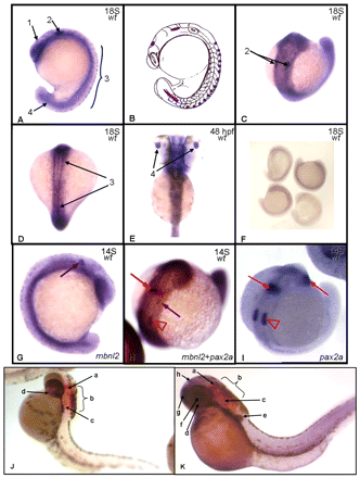
Expression pattern of mbnl2 in zebrafish. During the segmentation period (A–D), the embryos display an mbnl2-positive signal in mesencephalon (1), at hindbrain level (2), in spinal cord neurons (3) and in neural tube (4). Panel B shows a schematic representation of the embryo in panel A, with the mbnl2 signal symbolized in purple. (E) At 48 hpf, during the hatching period zebrafish mbnl2 is also expressed in the lens (4). (F) The sense control probe allows us to differentiate between background and the true signal. (G) Single detection hybridization (pax2a + mbnl2) and (I) single detection of pax2a transcripts in wild-type zebrafish at 14 somites. Red arrows signal the position of pax2a-positive cells at the midbrain-hindbrain boundary and immature eye; red arrowheads show the position of the otic vesicles; purple arrows signal mbnl2-expressing cells. (J) At 48 hpf, pax2a is expressed in the midbrain-hindbrain boundary (a), hindbrain (b), otic capsule (c) and optic nerve (d). (K) During the same stage, mbnl2 is expressed in the pectoral fin bud (e), lens (f), telencephalon (g) and epiphysis (h). The two signals do not overlap.