
|
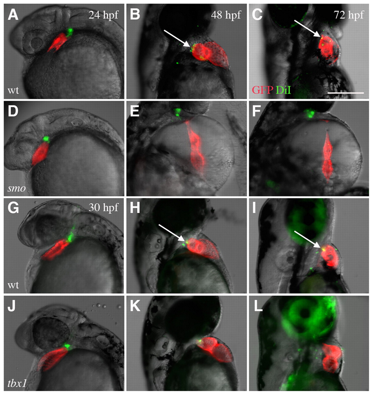
Fig. 9 Labeled cells in the pericardial wall are not incorporated into the developing heart in smo and tbx1 mutants. (A-C,G-I) Cells labeled with DiI (pseudocolored green) in the branchial region are incorporated into the myocardium (pseudocolored red) in wild-type zebrafish hearts after labeling at 24 hpf (A-C) and 30 hpf (G-I). DiI colocalized with the myocardium in wild-type fish at 48 and 72 hpf (arrows). (D-F,J-L) This incorporation of cells does not occur in most smo (D-F) or tbx1 (J-L) mutants. The green cells that appear to overlap with red cardiac cells (K) do not overlap with the myocardium 24 hours later (L). All embryos expressed cmlc2-GFP. Scale bar: 25 μm.