
|
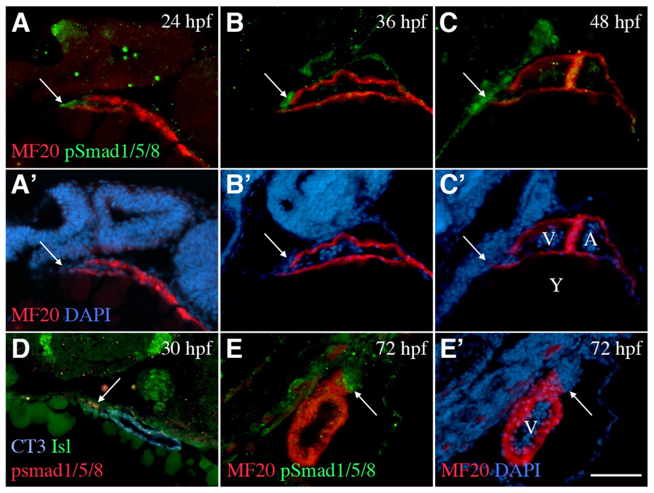
Fig. 6 Phosphorylated Smad1/5/8 is present in cells being incorporated into the arterial pole up to 72 hpf. Sagittal sections of wild-type zebrafish, cranial to the top, dorsal to the left. (A-C′) pSmad1/5/8 is detected in cells adjacent to and within the arterial pole (arrows) at 24, 36 and 48 hpf during addition of the second heart field myocardial cells to the heart tube (MF20). (D) pSmad1/5/8 (red) is expressed in a subset of Isl1-positive cells (green) closest to the myocardium (CT3) at 30 hpf. (E,E′) By 72 hpf, pSmad1/5/8 (green) is expressed in the myocardium and the smooth muscle (red) of the bulbus arteriosus. A, atrium; V, ventricle; Y, yolk. Scale bar: 5 μm.