
Image
|
Figure Caption
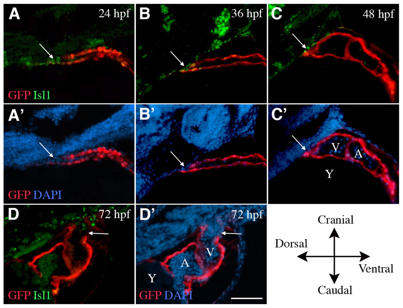
Fig. 5 Isl1 is expressed by cells that are being incorporated into the arterial pole up to 48 hpf. Sagittal sections of Tg(cmlc2::GFP) zebrafish, cranial to the top, dorsal to the left. (A-C′) Isl1 (green) is expressed in cells adjacent to and within the arterial pole (arrows) at 24, 36 and 48 hpf during addition of the second heart field myocardial cells to the heart tube (GFP, red). (D,D′) By 72 hpf, Isl1 is absent from the myocardium and is expressed in the cranial-most portion of the smooth muscle of the bulbus arteriosus. A, atrium; V, ventricle; Y, yolk. Scale bar: 5 μm.
Acknowledgments
This image is the copyrighted work of the attributed author or publisher, and
ZFIN has permission only to display this image to its users.
Additional permissions should be obtained from the applicable author or publisher of the image.
Full text @ Development