
|
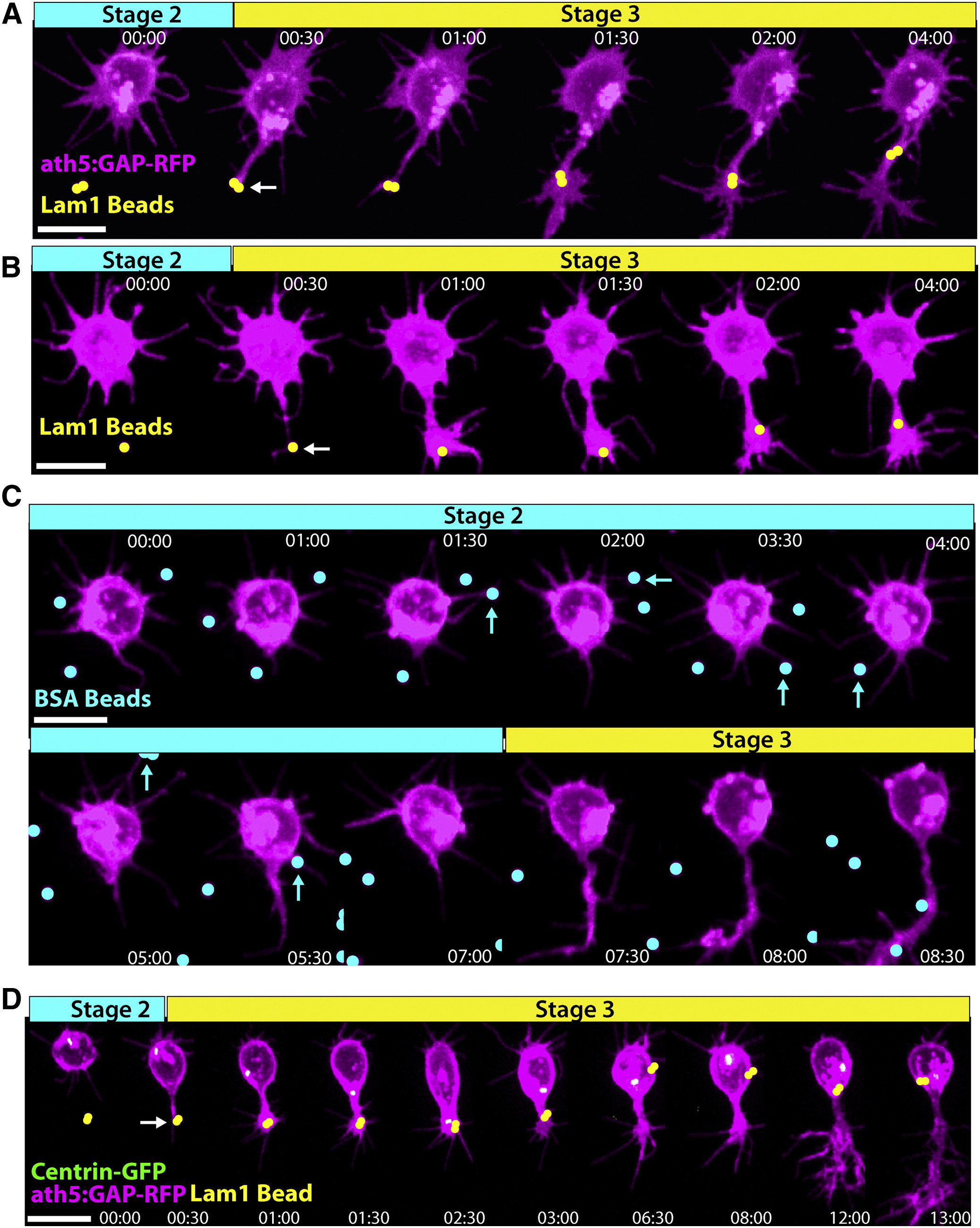
Fig. 5
Lam1 Contact Is Sufficient to Transform a Stage 2 Neurite into an Axon In Vitro
Dissociated RGCs from ath5:GAP-RFP embryos were plated on poly-L-lysine scattered with Lam1-coated 1 μM polystyrene beads (pseudocolored yellow) and analyzed by time-lapse microscopy.
(A and B) When a Stage 2 neurite contacts a Lam1 bead (contact point marked by arrow), this quickly (within one 30 min time point) induces a dramatic transformation from a thin neurite to one with an elaborate growth cone typical of an RGC axon.
(C) When presented with BSA-coated control beads (pseudocolored cyan), neurite contact (cyan arrows) does not have an observable effect.
(D) Imaging of cultured ath5:GAP-RFP/Centrin-GFP RGCs contacting a Lam1 bead (pseudocolored yellow, arrow indicates contact point) demonstrates that along with axon induction, neurite contact causes the centrosome to orient toward the site of Lam1 contact, and induces a transient migration toward the bead.
Frames are taken from [Movie S8. Contact with Lam1-Coated Beads Rapidly Transforms a Neurite into a Growth Cone-Tipped Axon In Vitro] and [Movie S9. Neurite Contact with Lam1 Causes Centrosome Reorientation and Somal Translocation toward Lam1 In Vitro, as well as Axon Induction]. Time is shown in hr:min; scale bars = 10 μM.