
|
Fig. 3
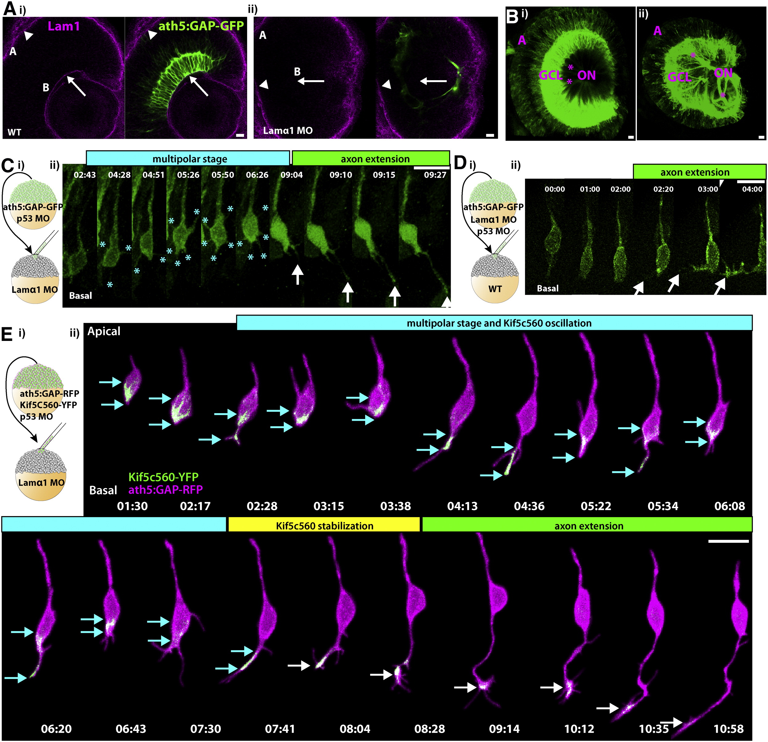
Lamα1 Is Necessary for Directed RGC Polarization
(Ai) Immunofluorescent staining of ath5:GAP-GFP embryos with polyclonal rabbit anti-Lam1 antibody reveals strong staining at the basal lamina lining the basal surface of the retina, or ILM (B, arrow), as well as at the basal lamina of the RPE, or Bruch′s membrane (A, arrowhead). (Aii) Injection of an antisense morpholino targeted against Lamα1 results in efficient loss of Lam1 staining at the basal surface (arrow), while Lam1 staining at Bruch′s membrane remains. Images are of a single confocal slice. (B) Confocal reconstruction from a WT 3 dpf retina (Bi) demonstrating the highly ordered nature of the ganglion cell layer (GCL) and the RGC axon fascicles (*) collecting to form the optic nerve (ON). (Bii) After lamα1 morpholino injection, the GCL is disorganized, as are the axon fascicles, which meander through the retina before colleting to form the ON. (Ci) Mosaic embryos with WT ath5:GAP-GFP-labeled RGCs in a lamα1 morphant environment were analyzed by time-lapse confocal microscopy beginning at approximately 35 hpf. (Cii) RGCs in this environment progress through a transient multipolar phase (marked by [*], cyan phase) before projecting an axon (arrow, green phase). (Di) Mosaic embryos with lamα1 morphant, ath5:GAP-GFP-labeled RGCs in a WT environment were analyzed by time-lapse confocal microscopy. (Dii) Morphant RGCs behave normally in this environment, and axons project directly from the basal surface of the cell (marked by arrowheads). (Ei) Mosaic embryos with WT ath5:GAP-GFP-labeled, Kif5c560-YFP-expressing RGCs in a lamα1 morphant environment were analyzed by time-lapse confocal microscopy. (Eii) In this context, Kif5c560-YFP signal accumulation (marked by cyan arrows) oscillates between the cell body and transient neurites (cyan phase) before stably accumulating in a single neurite (marked by white arrows, yellow phase) that extends to form the axon (green phase). Note that the individual confocal z-slices were cropped to remove signal not associated with the cell. A reconstruction of the uncropped frames is shown in Movie S5. Frames are taken from [Movie S3. Lamα1 Is Required for Highly Directed RGC Polarization], [Movie S4. Lamα1 Is Dispensable within RGCs for Highly Directed Polarization] and [Movie S5. In a Lamα1-Deficient Environment, Polarizing RGCs Exhibit Oscillations in Kif5c560-YFP Signal Accumulation Typical of Stage 2 Neurons Polarizing In Vitro]. Time is shown in hr:min; scale bars = 10 μM.