
|
Fig. 1
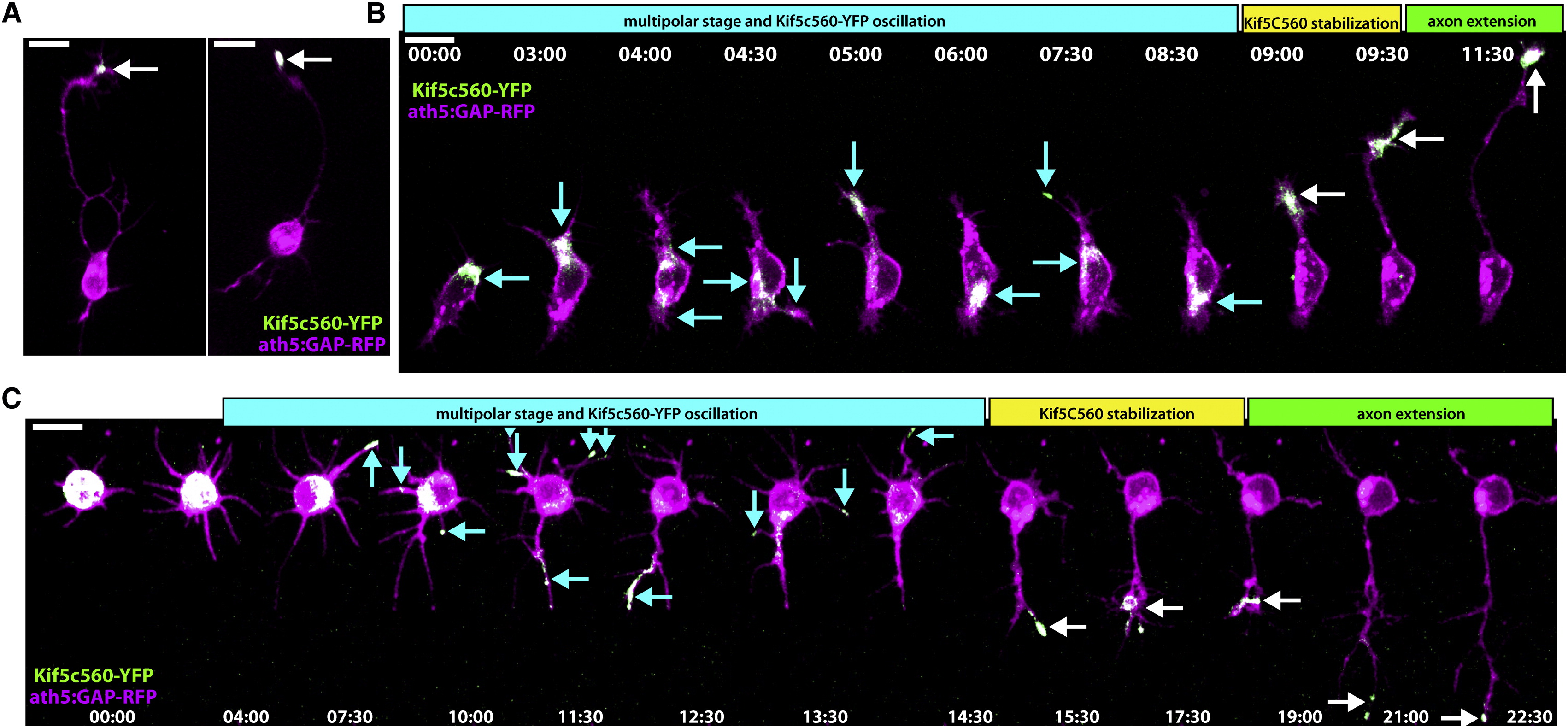
Kif5c560-YFP Marks the Axon of Cultured RGCs and Oscillates during Stage 2
Retinal ganglion cells (RGCs) were dissociated from ath5:GAP-RFP embryos injected with Kif5c560-YFP mRNA at the one-cell stage and allowed to polarize in vitro. (A) Bright Kif5c560-YFP was seen in the growth cone of the long axon projecting from polarized RGCs (arrow). (B and C) Shortly after plating, RGCs exhibit Kif5c560-YFP oscillations in different areas of the cell body and individual neurites, typical of Stage 2 behavior (arrows, cyan phase). The construct eventually stabilizes in a single neurite (white arrows, yellow phase), and this neurite extends to form the axon (green phase). Figure is related to Movie S1. Time is shown in hr:min; scale bars = 10 μM.