
|
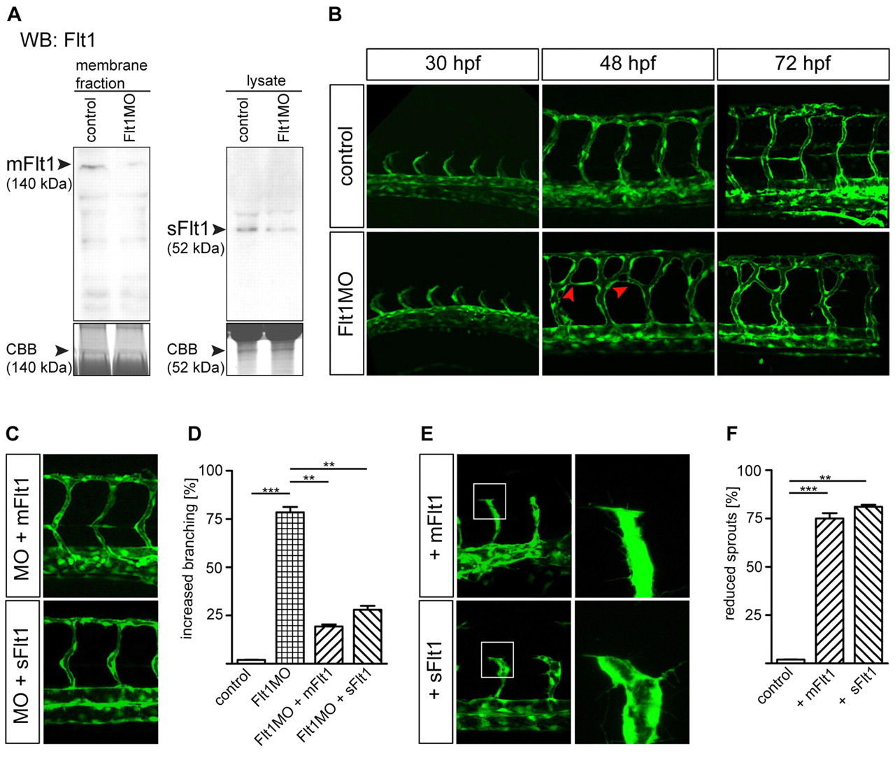
Fig. 2 Flt1 regulates segmental vessel branching morphogenesis. (A,B) Reduced mFlt1 and sFlt1 protein levels (A) and excessive segmental vessel branching (B) in zebrafish flt1 morphants. The red arrowheads in B indicate the aberrant connection between adjacent segmental vessels. (C,D) Injection of mRNA encoding mflt1 or sflt1 rescued segmental branching defects in flt1 morphants. MO, flt1 ATG morpholino. y-axis shows percentage of examined embryos with aberant segmental vessel branching, as shown in B (lower middle panel; 120 embryos/group from four separate experiments). (E,F) Overexpression of mflt1 (top row) or sflt1 (bottom row) in control embryos results in short segmental artery sprouts (E, left) and reduced filopodia extensions (E, right). The boxed regions are shown at higher magnification in the right-hand panels. y-axis shows percentage of embryos with reduced segmental vessel sprouts, as shown in E. **, P<0.01; ***, P<0.001; Student′s t-test. Error bars indicate s.e.m. (120 embryos/group from three separate experiments).