
|
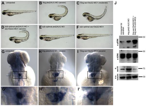
Fig. 4 Knockdown of sec10 partially phenocopies loss of pkd2.
(A-F) Gross phenotypes of zebrafish embryos at 3 dpf, lateral view, 4x magnification. Uninjected embryo (A), 4ng pkd2MO embryo with a severe curly tail up (B), 15ng sec10MO embryo with a moderate curly tail up (C). A synergistic interaction resulting in severe curly tail up was observed upon co-injection of sub-optimal doses of 0.25/2ng pkd2 MO +7.5ng sec10MO (D)—which do not result in curly tail up when injected alone (E, F). (G-I′) in situ hybridization for wt1a (with enlarged insets), 3 dpf, dorsal view, 16x magnification. An uninjected embryo with condensed glomerular stain (G/G′), a 4ng pkd2MO embryo with severe enlargement (H/H′), and a 15ng sec10MO embryo with severe enlargement (I/I′). (J) Increased phospho-ERK levels detected by Western blot in 4ng pkd2MO and 8+8ng sec10MO embryos at 5 dpf. One blot, loaded at 2 embryos per lane, was probed with antibody against phospho-ERK, then with antibody against total-ERK. The other blot, with the same lysates as above loaded at 10 embryos per lane, was probed with antibody against hSec10, then with antibody against gamma-tubulin.