
Image
|
Figure Caption
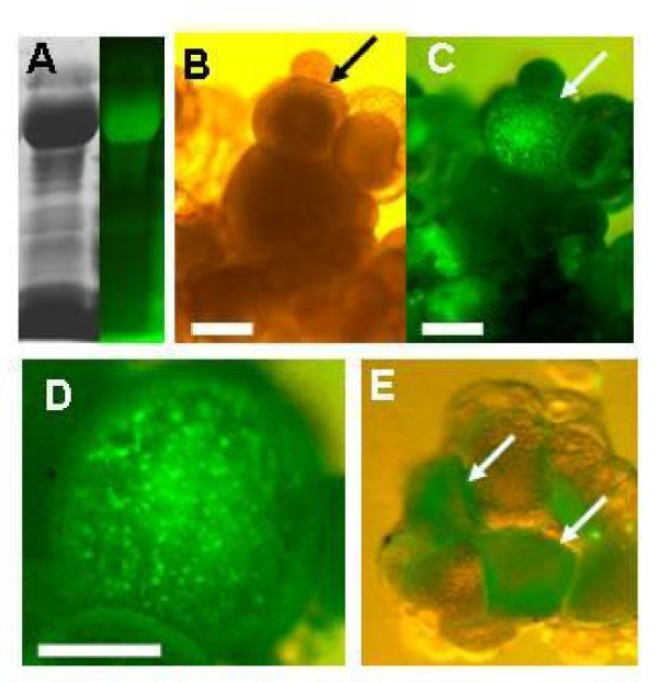
Fig. 5
In vitro incorporation of FITC-labeled Vtg derived from the bubble-eye goldfish by zebrafish oocytes. (A) Stability of FITC-labeled Vtg in the culture medium. The medium was subjected to SDS-PAGE after 48 h of incubation at 28°C. (B) Ovarian explant at 48 h of culture with FITC-labeled Vtg solution. (C) FITC fluorescence in the explant. (D) Enlargement of the ovary emitting FITC fluorescence. (E) Ovary explant cultured with purified FITC-labeled Vtg. Arrows show ovaries emitting FITC fluorescence. Scale bar = 200 μm.
Acknowledgments
This image is the copyrighted work of the attributed author or publisher, and
ZFIN has permission only to display this image to its users.
Additional permissions should be obtained from the applicable author or publisher of the image.
Full text @ Reprod. Biol. Endocrinol.