
Image
|
Figure Caption
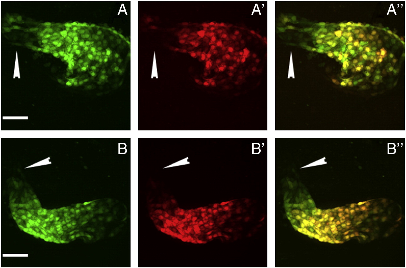
Fig. S3 Late myocardial addition occurred during a short developmental period. Hearts exposed to UV at (A–A′′) 23 hpf and imaged at 28 hpf or (B–B′′) at 28 hpf and imaged at 32 hpf. White arrowheads indicate green-only fluorescing cells. Green channel (A and B), red channel (A′ and B′), and overlay (A′′ and B′′). Scale bar represents 50 μm.
Acknowledgments
This image is the copyrighted work of the attributed author or publisher, and
ZFIN has permission only to display this image to its users.
Additional permissions should be obtained from the applicable author or publisher of the image.
Reprinted from Developmental Biology, 354(1), Lazic, S., and Scott, I.C., Mef2cb regulates late myocardial cell addition from a second heart field-like population of progenitors in zebrafish, 123-133, Copyright (2011) with permission from Elsevier. Full text @ Dev. Biol.