
|
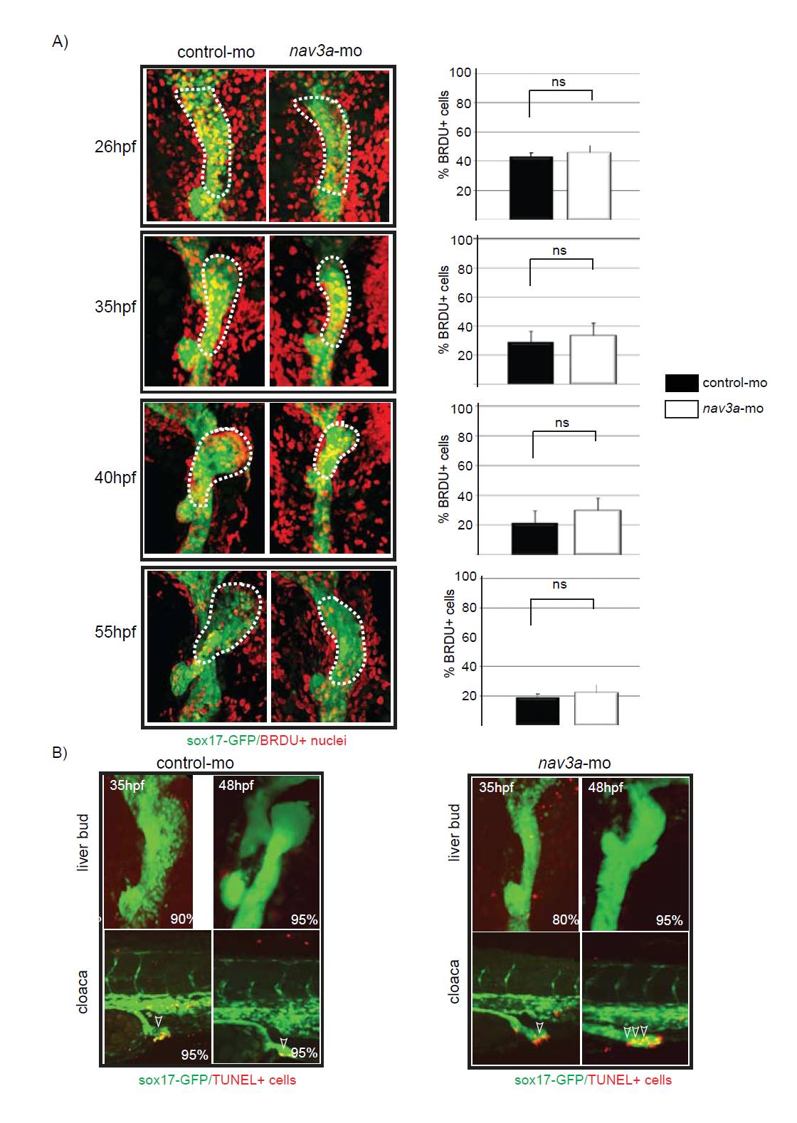
Fig. S5 Proliferation and apoptosis are not affected in the liver bud of nav3a morphant embryos. (A) Four hours before the indicated time points BrdU was injected in the yolk sac of control- and nav3a-morpholino-injected sox17:GFP embryos. After harvesting the embryos, BrdU labeling was performed. BrdU-positive cells between the pancreatic bud and the beginning of the pharyngeal endoderm were counted. No significant differences were observed in the relative amount of BrdU-positive cells at (n=5-7/group). Data are expressed as mean ± s.e.m. (B) Apoptosis was analyzed by TUNEL assay. No apoptosis could be detected in the liver bud of control or nav3a morphant embryos (upper panel). By contrast, increased amounts of TUNEL positive nuclei could be detected in intestinal endoderm of the cloaca region of nav3a morphant embryos (lower right panel). The percentage of embryos exhibiting a similar staining pattern is indicated in the lower right corner (n=20 embryos/group). A and B show ventral view.