
Image
|
Figure Caption
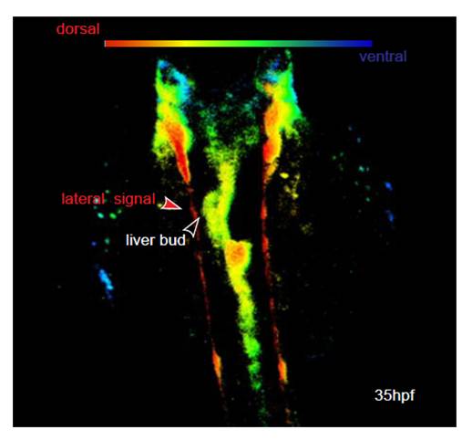
Fig. S11 Lateral GFP positive cells (as seen in Movies 1 and 2) are dorsal to the foregut endoderm and are not in contact to the liver bud. Sox17:GFPs870 embryos at 35 hpf imaged by confocal microscopy. Dorsolateral orientation of GFP positive cells in the embryo are indicated by color coding (red, dorsal; blue, ventral). The lateral signal in Movie 1 in the supplementary material is identical to the cells in the dorsal aspect. They are at the dorsal roof of the embryos and not in contact with the foregut endoderm.
Acknowledgments
This image is the copyrighted work of the attributed author or publisher, and
ZFIN has permission only to display this image to its users.
Additional permissions should be obtained from the applicable author or publisher of the image.
Full text @ Development