
|
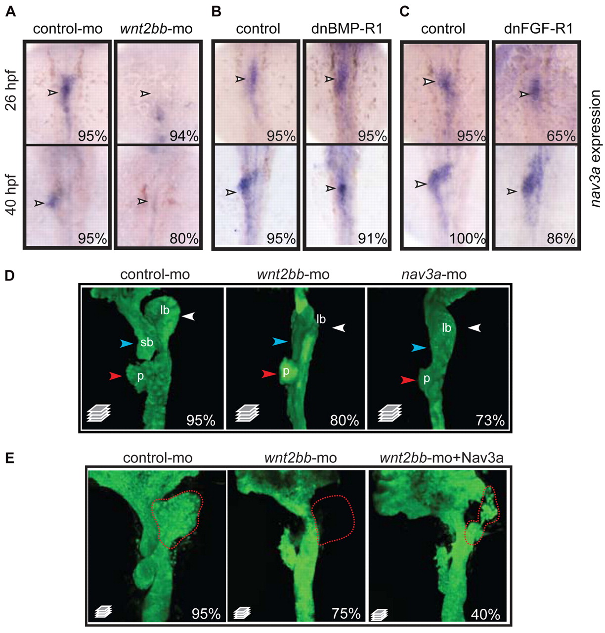
Fig. 5 Nav3a expression is controlled by Wnt2bb signaling. (A) Embryos injected with control morpholino or wnt2bb morpholino examined for nav3a expression by in situ hybridization. Wnt2bb inhibition results in reduced nav3a expression in the liver bud (arrowheads). (B,C) Embryos obtained from outcrossing of hemizygous Tg(hsp70l:dnBmpr-GFP) or Tg(hsp70l:dnfgfr1-GFP) zebrafish were heat shocked at 18 hpf, collected at 40 hpf and examined for nav3a expression by in situ hybridization. Inhibition of BMP and FGF receptor signaling did not annihilate nav3a expression (arrowheads) in the liver bud. (D) Wnt2bb depletion phenocopies nav3a depletion. Sox17:gfp embryos injected either with control morpholino (left-hand panel), wnt2bb morpholino (middle panel) or nav3a-ATG morpholino (right-hand panel) examined at 40 hpf by confocal microscopy. Both wnt2bb- and nav3a-morphant embryos developed an aberrant liver bud (white arrowhead). Also the pancreatic bud and the swim bladder primordium did not develop properly (red and blue arrowheads, respectively). (E) Endodermal nav3a overexpression partially rescued wnt2bb inhibition. Sox17-gfp embryos were injected with control morpholino, wnt2bb morpholino alone or wnt2bb morpholino together with sox17-nav3a-mcherry plasmid and analyzed at 50 hpf. Wnt2bb inhibition induced aberrant development of the liver bud, pancreatic bud and swim bladder primordium (middle panel). In 40% of embryos, endodermal nav3a overexpression results in a partial rescue of liver bud formation (right-hand panel). Nevertheless these liver buds appeared to be aberrantly shaped (right-hand panel). Red dotted lines indicate the liver bud. The percentage of embryos exhibiting a similar phenotype as displayed in the figure, is indicated in the lower right-hand corner (A-C: n=10-30 embryos per group; D: n=150 per group, E; n=100 per group). lb, liver bud; sb, swim bladder primordium; p, pancreatic bud. A-C show dorsal view; D and E show ventral view.