
IMAGE
Fig. s4
- ID
- ZDB-IMAGE-110510-9
- Publication
- Lobbardi et al., 2011 - Fine-tuning of Hh signaling by the RNA-binding protein Quaking to control muscle development
- All Figures
- Figures for Lobbardi et al., 2011
Image
|
Figure Caption
Fig. s4
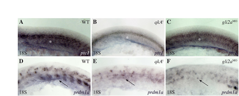
Reduction of the Hh signaling pathway at the 18-somite stage in qkAt and gli2aMO. (A-F) qkAt and gli2aMO embryos exhibit hallmarks (white asterisks and black arrows point to the adaxial cell derivatives labeled by ptc1 and prdm1a, respectively) of downregulated Hh signaling. Embryos were fixed at the 18-somite stage and processed for in situ hybridization with ptc1 (A-C) and prdm1a (D-F) probes. Lateral views, anterior to the left.
Acknowledgments
This image is the copyrighted work of the attributed author or publisher, and
ZFIN has permission only to display this image to its users.
Additional permissions should be obtained from the applicable author or publisher of the image.
Full text @ Development