
Fig. s6
- ID
- ZDB-IMAGE-110510-11
- Publication
- Lobbardi et al., 2011 - Fine-tuning of Hh signaling by the RNA-binding protein Quaking to control muscle development
- All Figures
- Figures for Lobbardi et al., 2011
|
Fig. s6
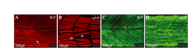
Partial restoration of the fast muscle cell phenotype at 48 hpf. (A-D) At 48 hpf, the slow muscle phenotype is not rescued in the mutant (B), which exhibits gaps (white asterisk) compared with WT embryos (A). The fast muscle fibers, constituting the second major muscle cell type, are organized in WT (C) in a stereotyped manner (diagonal direction of fibers, yellow arrowhead). At 48 hpf, in the qkAt mutant (D), the fast muscle fibers exhibit a myotube structure but are not as well organized as in the WT sibling (C, yellow arrowhead), indicating a partial rescue of the fast muscle fiber organization. Dorsal views, anterior to the left.