
Fig. s2
- ID
- ZDB-IMAGE-110411-1
- Publication
- Kikuchi et al., 2011 - Retinoic Acid production by endocardium and epicardium is an injury response essential for zebrafish heart regeneration
- All Figures
- Figures for Kikuchi et al., 2011
|
Fig. s2
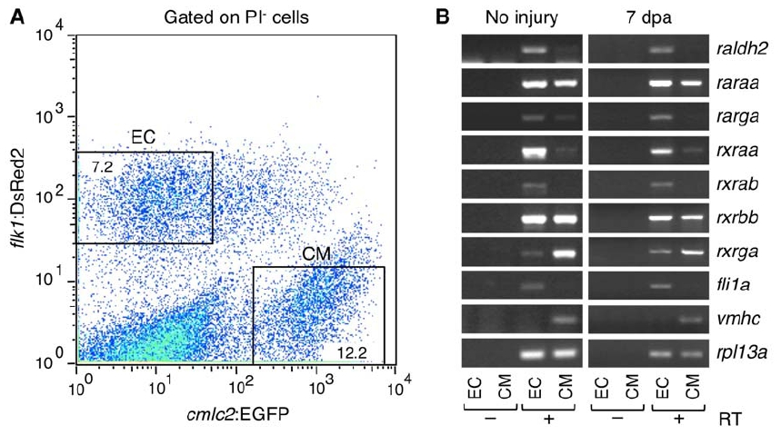
Expression Analyses of RA Signaling Components
(A) Endocardial cells (EC) and cardiomyocytes (CM) were purified by FACS from dissociated uninjured or 7 dpa cmlc2:EGFP; flk1:DsRed2 ventricles. A representative plot from 7 dpa ventricles is shown. Cells stained by propidium iodide (PI) are excluded as dead cells. Percentages of the total PI-negative cell population that were gated are shown.
(B) Expression of RA signaling components was examined by RT-PCR in purified endocardial cells (EC) and cardiomyocytes (CM). Endocardial (fli1a) and cardiomyocyte markers (vmhc) were used to confirm the specificity of cell sorting. ribosomal protein L13a (rpl13a), was used as a control. The amplified products were not detectable without reverse transcriptase (RT), confirming that bands are amplified from cDNAs of the target genes but not from genomic DNA contaminants. raldh2 transcripts detected in samples of uninjured ventricles are likely accentuated by the severe trauma of ventricular cell extraction and dissociation.
Reprinted from Developmental Cell, 20(3), Kikuchi, K., Holdway, J.E., Major, R.J., Blum, N., Dahn, R.D., Begemann, G., and Poss, K.D., Retinoic Acid production by endocardium and epicardium is an injury response essential for zebrafish heart regeneration, 397-404, Copyright (2011) with permission from Elsevier. Full text @ Dev. Cell