
|
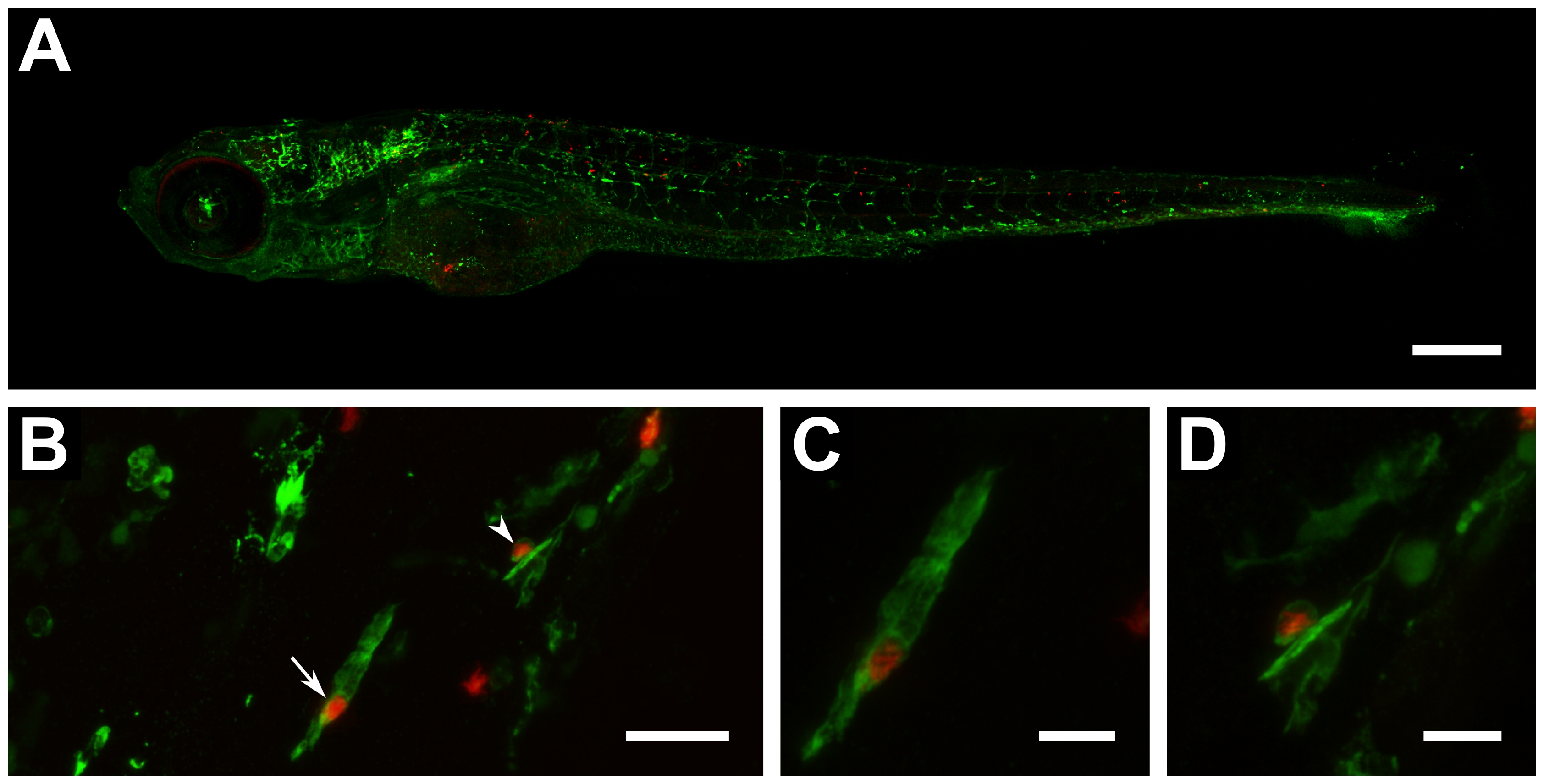
Fig. 6
Automatic yolk sac injection of M. tuberculosis and effect of treatment on infected larvae.
(A) Confocal z-stack (8×2 stitching) of a 6 day-old whole larva (fli1-egfp with gfp-labelled vasculature) showing spread of bacteria (red) throughout the body (scale bar: 250 μm). (B) Confocal z-stack of red-fluorescent bacteria co-localizing with green-fluorescent leukocytes detected by L-plastin immunostaining (scale bar: 25 μm). (C) Close-up (digital zoom: 4.2) of bacteria-containing leukocyte depicted in B by straight arrow (scale bar: 10 μm). (D) Close-up (digital zoom: 4.3) of bacteria-containing leukocyte depicted in B by arrowhead (scale bar: 10 μm).