
|
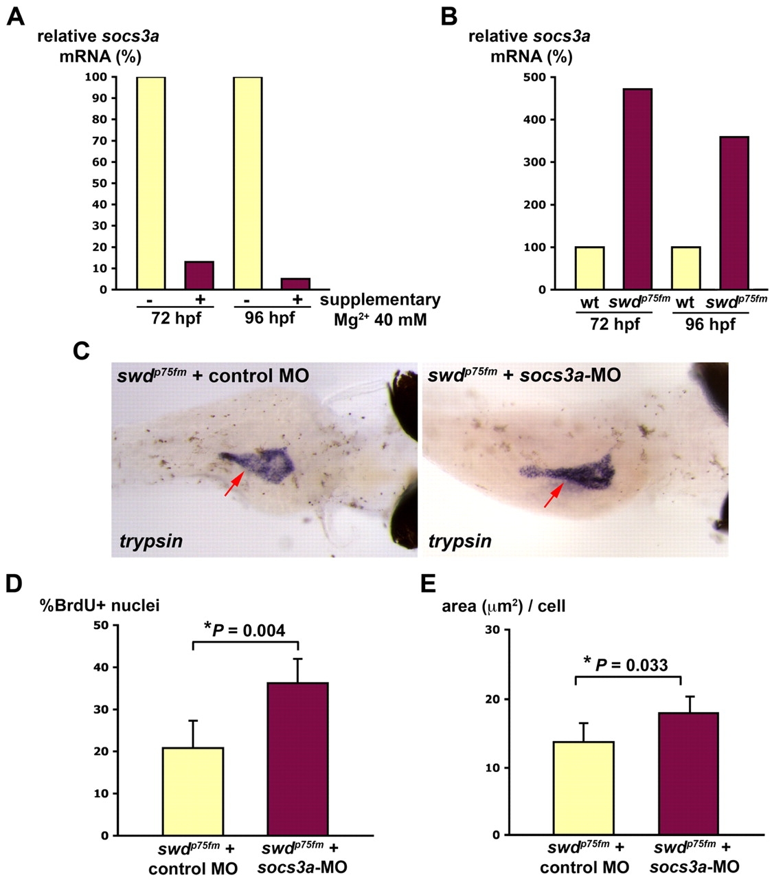
Fig. 6
The Mg2+-sensitive Socs3a pathway is involved in the proliferative defect of the swd mutants. Supplementary Mg2+ improves the exocrine pancreas phenotype of the swd mutants that have repression of socs3a. (A) swdp75fm mutants were incubated in E3 medium with or without supplementary 40 mM MgCl2 for 72 or 96 hpf and analyzed for socs3a mRNA by real-time PCR. (B) swdp75fm mutants and WT were incubated in E3 medium (containing 1.65 mM Mg2+) for 72 or 96 hpf, and the socs3a mRNA levels were determined by real-time PCR. (C) Embryos collected from swdp75fm/+ intercross were microinjected with socs3a-ATG-MO or control MO, incubated until 4 dpf and the exocrine pancreas (red arrows) was analyzed by whole-mount in situ hybridization using anti-trypsin riboprobes. The image is representative of 20 larvae in each group from three independent experiments. The swdp75fm mutants injected with either non-targeting control MO or socs3a-5-mispair MO are indistinguishable in the exocrine pancreas by in situ hybridization using anti-trypsin riboprobes. (D) socs3a-ATG-MO- or control-MO-injected larvae were incubated until 72 hpf, injected with BrdU and then analyzed for the proportion of cells in S phase (% BrdU+ nuclei). (E) socs3a-ATG-MO- or control-MO-injected larvae were incubated until 96 hpf and analyzed for cell growth (area, in μm2, per cell). *P<0.05 indicates statistically significant difference. (C-E) The swdp75fm mutants were identified on the basis of their hypopigmented skin, which was not grossly affected by socs3a-ATG-MO.