
Image
|
Figure Caption
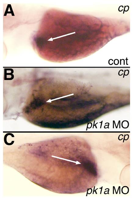
Fig. S7 Ceruloplasmin (cp) staining of 4 dpf larvae injected with pk1a MO. (A) In situ hybridization of 4 dpf larva injected with control (cont) MO demonstrating liver staining (white arrow). (B-C) In situ hybridizations of 4 dpf larvae injected with pk1a MO demonstrating liver staining (white arrow) similar to (A). Views in A and B are left lateral, while that in C is right lateral, showing the liver on the right side, similar to other studies depicted here.
Acknowledgments
This image is the copyrighted work of the attributed author or publisher, and
ZFIN has permission only to display this image to its users.
Additional permissions should be obtained from the applicable author or publisher of the image.
Reprinted from Developmental Biology, 351(2), Cui, S., Capecci, L.M., and Matthews, R.P., Disruption of planar cell polarity activity leads to developmental biliary defects, 229-241, Copyright (2011) with permission from Elsevier. Full text @ Dev. Biol.