
|
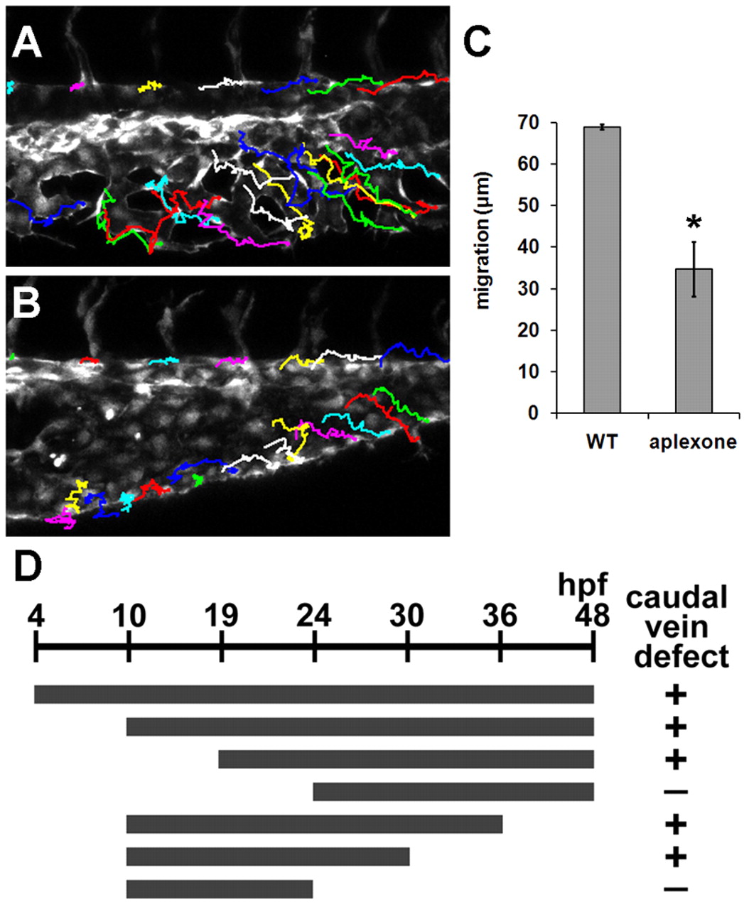
Fig. 3 Aplexone affects the migration of caudal vein endothelial cells. (A,B) Representative images of the caudal vein of control (A) and 10 μM aplexone-treated (B) embryos at 30 hpf. Images were overlaid with the endothelial cell migration path inferred from time-lapse confocal movies taken from 25 hpf to 30 hpf. The migration of caudal vein endothelial cells was traced using the Manual Tracking feature of ImageJ and the positions of ISV sprouting points were used as references to adjust for the growth of embryos. (C) The distance of endothelial cell migration in caudal vein of control and aplexone-treated Tg(kdrl:GFP) embryos. Asterisk indicates P<0.05. (D) The window of application of aplexone. Gray bars represent the developmental stages during which Tg(kdrl:GFP) embryos were exposed to 10 μM aplexone and the caudal vein phenotype was analyzed at 48 hpf. + indicates that caudal vein angiogenesis was inhibited. – indicates the formation of normal caudal vein plexus.