
|
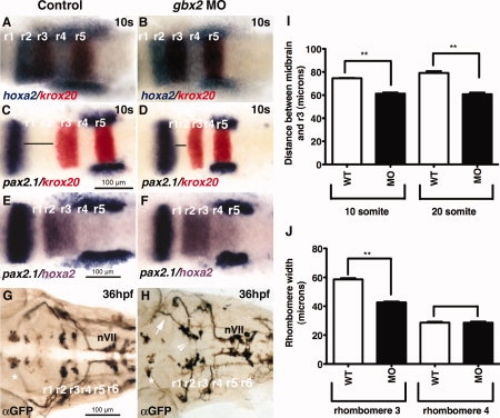
Fig. 1
gbx2 morpholino decreases the distance between midbrain and r3 and disrupts cranial nerve V motor neuron organization. A–F: Dorsal views of wild-type and gbx2 morphant anterior hindbrains. A,B: Two-color in situ hybridization examining the expression of hoxa2 (blue) and krox20 (red) at 10-somite stage. C,D: Two-color in situ hybridization examining the expression of pax2.1 (black) and krox20 (red) at 10-somite stage. Anterior hindbrain truncation between posterior midbrain (pax2.1) and r3 (krox20) is visible in embryos injected with 8 ng gbx2-MO at 10-somites (D). E,F: Two-color in situ hybridization examining the expression of pax2.1 (black) and hoxa2 (purple) at 10-somite stage. G,H: Anti-GFP antibody staining at 36 hpf. Embryos that were injected with 8 ng of gbx2-MO show abnormal clustering of nV cell bodies clustering in r2 and r3 (arrowhead in H), and ectopic axonal projections (arrow in H) The migration and organization of nVII motor neurons in r4–r7 are not affected (G,H). I: Distance between posterior midbrain and r3. J: Rhombomere width. *Midbrain (nIII and nIV) motor neurons. **P < 0.0001 (Student′s t-test). hpf, hours post fertilization; MO, morpholino; r, rhombomere; s, somite stage.