
Image
|
Figure Caption
Fig. S8
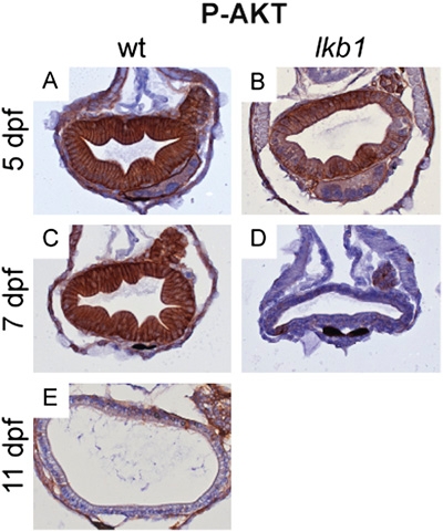
Deregulation of PI3K signaling in lkb1 mutants. Transverse sections of WT and lkb1 intestines at indicated days of development were stained with an antibody against phosphorylated AKT. (A and B) Strong phospho-AKT staining is detected in WT and lkb1-mutant intestines at 5 dpf. WT intestine at 7 dpf is strongly stained (C), whereas in lkb1-mutant intestine at 7 dpf phospho-AKT staining is barely detectable (D). (E) Starved WT larvae at 11 dpf show very little phospho-AKT staining in the intestine.
Acknowledgments
This image is the copyrighted work of the attributed author or publisher, and
ZFIN has permission only to display this image to its users.
Additional permissions should be obtained from the applicable author or publisher of the image.
Full text @ Proc. Natl. Acad. Sci. USA