
Image
|
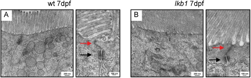
Figure Caption
Fig. S3
Intact apical brush border and tight junctions in lkb1-mutant intestines. Transmission electron microscope micrographs of WT and lkb1-mutant intestinal cells at 7 dpf. (A) The microvilli constituting the apical brush border are depicted in the intestinal cells. (B) Lkb1-mutant intestinal cells also have microvilli and an apical brush border in their apical surface. Magnifications show hemidesmosomes (black arrows) and tight junctions (red arrows) in WT and lkb1-mutant intestinal cells.
Acknowledgments
This image is the copyrighted work of the attributed author or publisher, and
ZFIN has permission only to display this image to its users.
Additional permissions should be obtained from the applicable author or publisher of the image.
Full text @ Proc. Natl. Acad. Sci. USA