
Image
|
Figure Caption
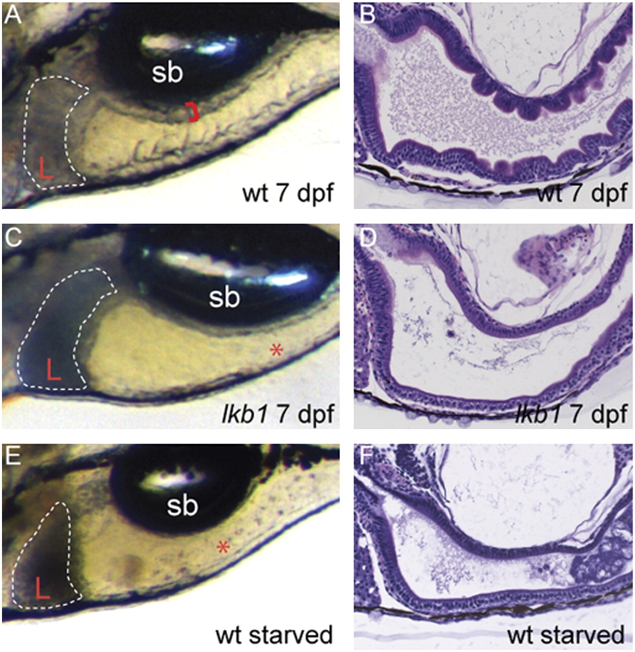
Fig. 2
The intestinal architecture of lkb1 mutants resembles that of starved WT larvae. (A, C, and E) High-power images depicting the liver and intestine of live larvae of the indicated genotypes; anterior is to the left. Red brackets demarcate the thickness of the intestinal wall, and livers are outlined. At 7 dpf, lkb1-mutant larvae (C) exhibit a small, dark liver (L) and flattened intestine (asterisk), as do starved 11-dpf WT larvae (E). (B, D, and F) H&E staining of sagittal sections of the intestine. Note loss of intestinal folding in 7-dpf lkb1- mutant larvae and in starved WT larvae. Sb, swim bladder.
Figure Data
Acknowledgments
This image is the copyrighted work of the attributed author or publisher, and
ZFIN has permission only to display this image to its users.
Additional permissions should be obtained from the applicable author or publisher of the image.
Full text @ Proc. Natl. Acad. Sci. USA