
|
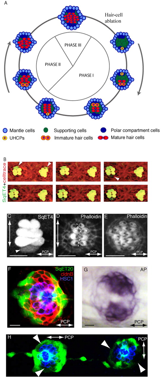
Fig. 1 Epithelial planar polarization of neuromasts. (A) Schematic description of the three phases of regeneration and homeostasis and the planar polarization of the neuromast epithelium (macula). Cell types are color-coded: most peripheral mantle cells (light blue), central supporting (sustentacular) cells (green), polar compartment resident cells (dark blue), UHCPs (yellow), immature hair cells (orange) and mature hair cells (red). The axis of planar polarity is evidenced by the position of the kinocilium within the mature hair cells (black dot). (B) Time-lapse of two adjacent parallel SqET4 neuromasts showing hair cells and their progenitors (green) and stained with Celltrace to reveal cellular boundaries (red). White arrowheads on the first panel indicate a pair of newly developed hair cells on the dorsum of both neuromasts. The white arrowhead on the third panel indicates a new ventral UHCP, which divides into two hair cells on the last panel. Hair cells develop in pairs, sequentially, and on the dorsal or ventral aspect of the neuromasts. (C) A regenerating SqET4 neuromast revealing six GFP-positive hair cells. A UHCP develops at the lower edge of the neuromast (arrowhead). The orthogonal axes of planar polarization (horizontal) and direction of regeneration (vertical) are indicated by double-headed arrows. (D) Actin staining of a regenerating neuromast, showing the orientation of four pairs of hair bundles and revealing the vertical line of bilateral symmetry (dotted white line). (E) Actin staining of neuromast in Phase III of regeneration showing the lateral expansion of the macula. A double-headed arrow indicates the axis of planar polarity. (F) A SqET20 neuromast expressing GFP (green) and stained with antibodies to Claudin-b (red), which marks all the supporting cells, and to HCS1 (blue), which highlights the hair cells. GFPlow compartments are located at the poles of the vertical axis of bilateral symmetry. (G) The GFPlow compartments coincide with the endogenous expression of alkaline phosphatase. In all figures, anterior is leftwards and dorsal is upwards. (H) A SqET20 larva expressing GFP (green) and stained with the antibody HCS1 (blue). The neuromast to the left of the image is L1 (a parallel neuromast), whereas the one on the right is L2 (a perpendicular neuromast). This image shows that the GFPlow compartments rotate through 90° in parallel versus perpendicular neuromasts. Arrowheads indicate polar compartments. Scale bars: 10 μm.