
|
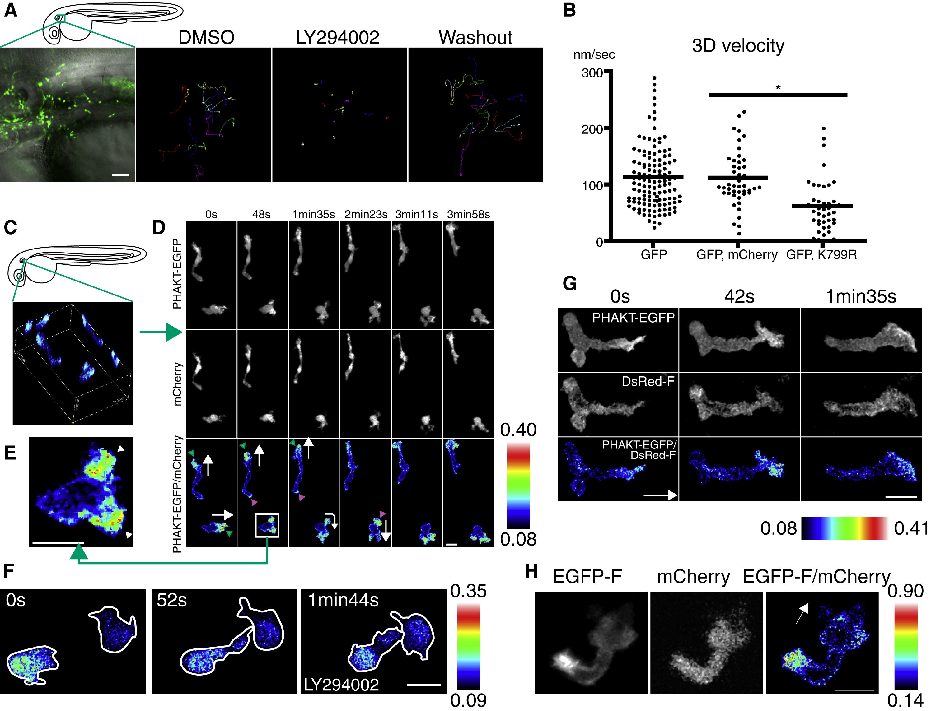
Fig. 3 PI(3)K Is Critical for Neutrophil Motility and Is Active at the Leading Edge in the Mesenchymal Tissues of the Head
(A) Random migration of neutrophils is arrested by 65 μM LY294002 and restored after washout of the drug. The lines indicate tracking of neutrophil motility (12 cells per condition) imaged for 30 min using Tg(MPO:GFP)uw (Movie S3A). Scale bar = 50 μm.
(B) PI(3)K γ K799R disturbs interstitial motility of neutrophils (Movie S3B, *p < 0.001, two-tailed unpaired t test; GFP: 128 neutrophils [40 movies], GFP, mCherry: 45 neutrophils [9 movies], GFP, K799R: 44 neutrophils [34 movies]).
(C) Three-dimensional reconstruction of ratiometric image (PHAKT-EGFP/mCherry).
(D) Time-lapse ratiometric imaging (PHAKT-EGFP/mCherry) of PI(3,4,5)P3-PI(3,4)P2 dynamics during random migration (Movie S4A). PI(3,4,5)P3-PI(3,4)P2 is mainly localized at the leading edge (green arrowheads) and occasionally at the tail (magenta arrow heads). White arrows indicate direction of migration. Scale bars in (D–H) = 10 μm.
(E) PI(3,4,5)P3-PI(3,4)P2 at the bifurcated pseudopod, indicated by arrowheads (Movie S4A).
(F) Treatment with 65 μM LY294002 inhibits the leading edge signal of PI(3,4,5)P3-PI(3,4)P2 and induces high ratiometric signals of PHAKT-EGFP/mCherry in the cell body of neutrophils (Movie S4C). Note the rounded tails and thin pseudopods induced by LY294002.
(G) PI(3,4,5)P3-PI(3,4)P2 signal at the leading edge by ratiometric imaging of PHAKT-EGFP/farnesylated DsRed (DsRed-F) (Movie S4D). The white arrow indicates direction of migration.
(H) Ratiometric imaging of EGFP-F/mCherry reveals periodic accumulation of membrane components at the tail (Movie S4E). The white arrow indicates direction of migration. Images are representative of three (A) and more than five (C–H) time-lapse movies from a minimum of three separate experiments.
Reprinted from Developmental Cell, 18(2), Yoo, S.K., Deng, Q., Cavnar, P.J., Wu, Y.I., Hahn, K.M., and Huttenlocher, A., Differential Regulation of Protrusion and Polarity by PI(3)K during Neutrophil Motility in Live Zebrafish, 226-236, Copyright (2010) with permission from Elsevier. Full text @ Dev. Cell