
|
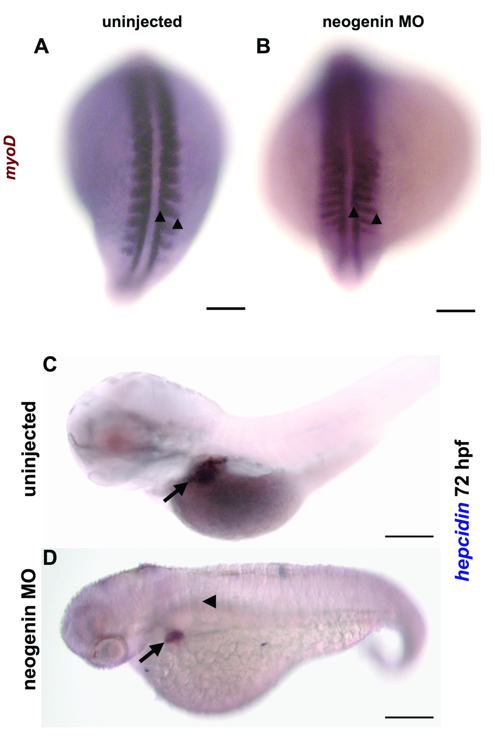
Fig. S4 Neogenin knockdown reproduced the reported defect in somitogenesis associated with neogenin deficiency. A,B. Whole mount in situ hybridization for myoD to stain the somites in uninjected (A) and neogenin morphants (B) at the 20 somites′ stage of development (dorsal view) confirmed that injection of the neogenin morpholino at 0.15 mM produced elongation of the somites, manifest by increased distance between the two arrowheads. This is characteristic of the neogenin deficient phenotype, as described by [4]. Scale bar represents 100 microns. C,D. Whole mount in situ hybridization for hepcidin at 72 hpf in uninjected control embryos (C) and neogenin morphants (D) (lateral view) revealed a shortened body axis with a curved tail and flattened somites (arrowhead) in the neogenin morphants. Hepcidin expression is present in the liver (arrow) of the neogenin morphant, although the expression domain of hepcidin is smaller than in the uninjected control. Scale bar represents 200 microns. N = 20 embryos per group. Embryos were photographed at 100x magnification with a an Axio Imager 1 compound microscope (Carl Zeiss MicroImaging, Inc., Thornwood, NY) and an AxioCam ICc1 digital camera (Carl Zeiss MicroImaging, Inc.) (A,B) or a BX51 compound microscope (Olympus, Center Valley, PA) and a Q-capture 5 digital camera (QImaging, Surrey, BC, Canada) (C,D).