
|
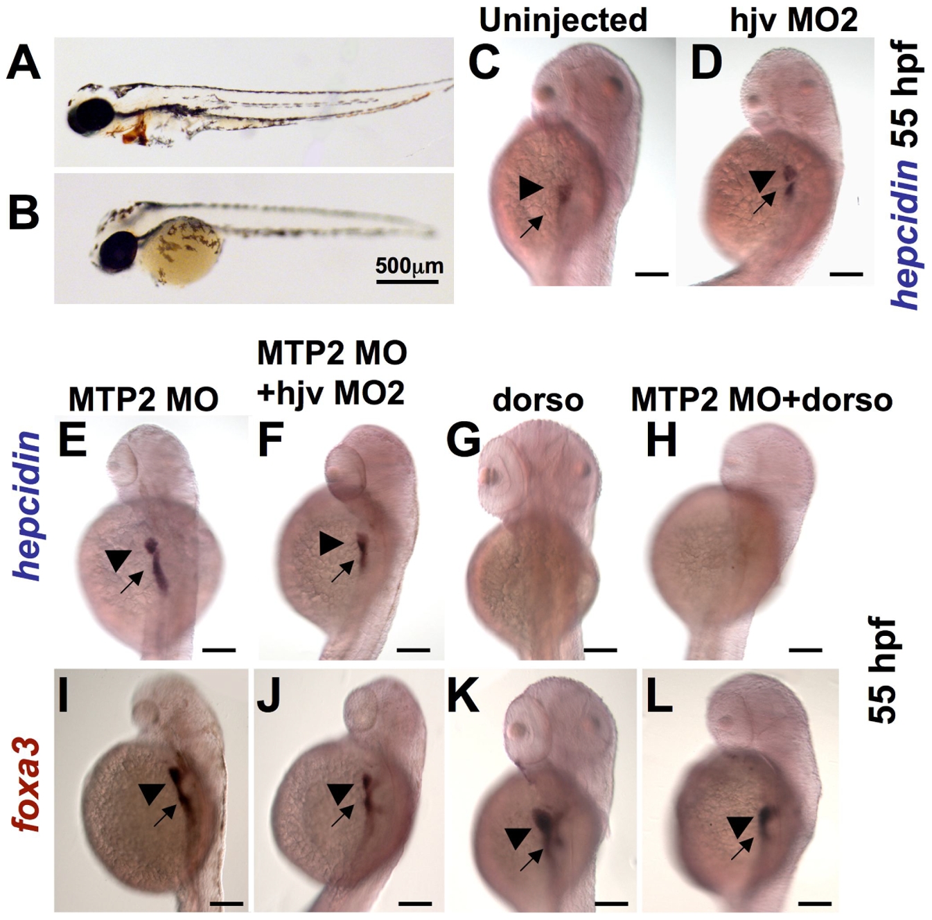
Fig. 6 Knockdown of mtp2 enhances expression of hepcidin at 55 hpf.
A–B. O-dianisidine staining for hemoglobin at 48 hpf demonstrated normal levels of hemoglobin in the cardiac circulation of WT embryos (A), but decreased hemoglobin in the mtp2 morphants (B). C–H. Whole mount in situ hybridization for hepcidin at 55 hpf demonstrated normal staining in uninjected controls (C) and hjv morphants (D). Knockdown of mtp2 (E) caused developmental delay, but increased the intensity of hepcidin staining in the liver (arrowhead) and the extent and intensity of staining in the intestine (arrow). Co-injection of hjv MO and mtp2 MO (F) resulted in a smaller embryo, but preserved hepcidin staining in the liver and intestine. Treatment with dorsomorphin from 28–55 hpf abrogated hepcidin expression in both uninjected embryos (G) and mtp2 morphants (H). I–L. Whole mount in situ hybridization for foxa3 demonstrated smaller liver size (arrowhead) in embryos injected with mtp2 MO (I,J,L), compared to dorsomorphin alone (K) or untreated embryos (compare with Figure 3D). N = 20–30 embryos per group.