
Image
|
Figure Caption
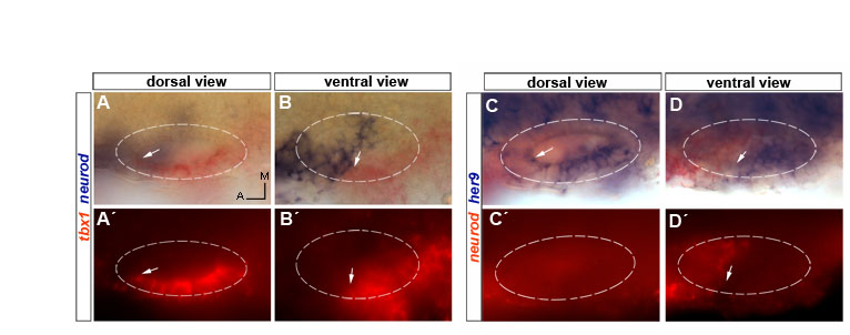
Fig. S1 Complementary expression of neurod and tbx1/her9 in 24 hpf wild-type embryos. (A-B′) Double in situ hybridization for neurod (blue) and tbx1 (red) at two different focal planes. Arrows point to the anterior limit of tbx1 expression. (C-D′) Double in situ hybridization for neurod (red) and her9 (blue) at two different focal planes. Arrows point to the anterior limit of her9 expression (C,D) and to posterior limit of neurod expression (D′).
Figure Data
Acknowledgments
This image is the copyrighted work of the attributed author or publisher, and
ZFIN has permission only to display this image to its users.
Additional permissions should be obtained from the applicable author or publisher of the image.
Full text @ Development