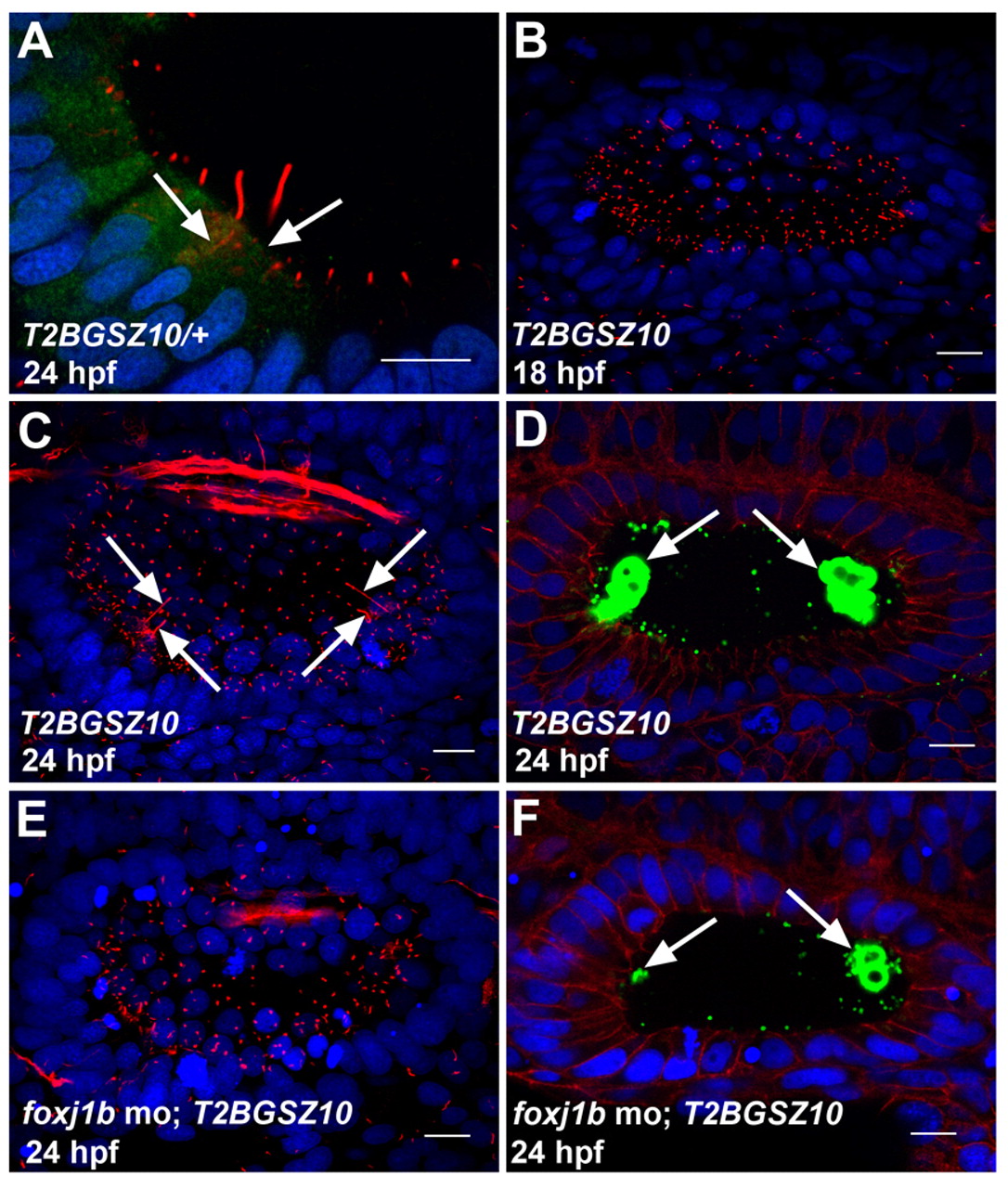
Fig. 4 Foxj1b is required for the differentiation of motile and kinocilia. (A) Expression of GFP in the hair cells of a heterozygous T2BGSZ10 transgenic zebrafish embryo. Arrows indicate hair cells. (B) Defective motile cilia differentiation in the ear of a homozygous T2BGSZ10 transgenic embryo (compare with Fig. 1E). (C) Defective kinocilia differentiation in the ear of a homozygous T2BGSZ10 transgenic embryo. Note the variable lengths of the kinocilia (arrows) (compare with Fig. 1F). (D) Malformed otoliths (green, arrows) in the ear of a homozygous T2BGSZ10 transgenic embryo. (E) A foxj1b morpholino-injected homozygous T2BGSZ10 transgenic embryo showing a more pronounced effect on ciliary differentiation with severe reduction in the motile and kinocilia. (F) A more prominent effect on otolith (green, arrows) formation in the ear of a foxj1b morpholino-injected homozygous T2BGSZ10 transgenic embryo. Embryos in A-C,E were stained with anti-acetylated tubulin antibodies (red), and those in D and F with anti-Stm antibodies (green). The embryo in A was co-stained with anti-GFP antibodies (green), whereas those in D and F were co-stained with anti-β-catenin antibodies (red). Nuclei were visualized with DAPI (blue). All panels show lateral views of otic vesicles with anterior to the left. Scale bars: 10 μm.
Image
Figure Caption
Figure Data
Acknowledgments
This image is the copyrighted work of the attributed author or publisher, and
ZFIN has permission only to display this image to its users.
Additional permissions should be obtained from the applicable author or publisher of the image.
Full text @ Development

