
|
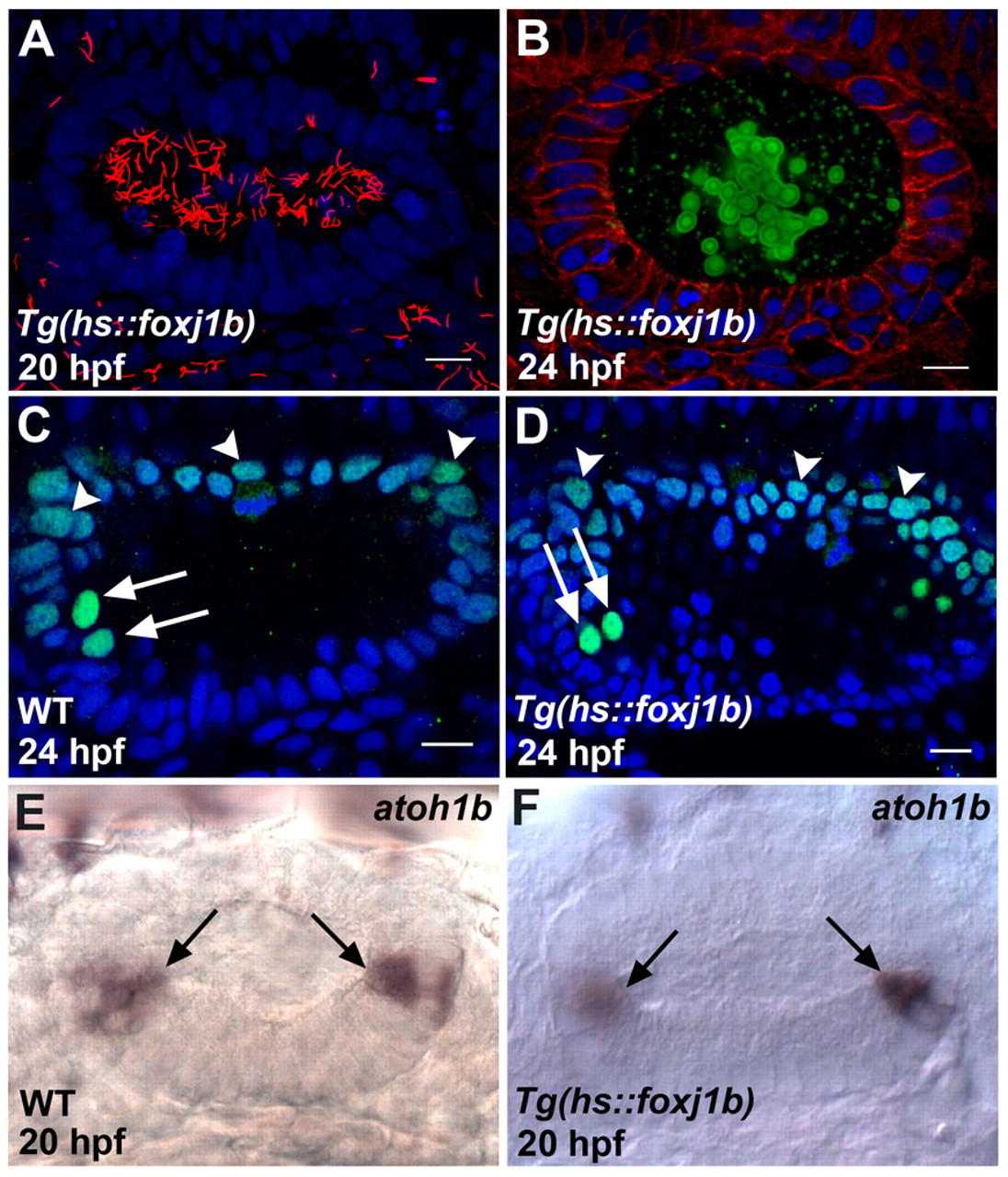
Fig. 3 Proper otolith formation requires a specific spatiotemporal distribution of motile cilia. (A) Ear of a zebrafish embryo overexpressing foxj1b, with large numbers of long motile cilia (red) throughout the otic vesicle. (B) Ear of a foxj1b-overexpressing embryo showing defective otolith (green) formation. (C) Wild-type pattern of Pax2 expression (green) in the dorsal part of the otic vesicle (arrowheads) and in the hair cells (arrows). The two anterior hair cell nuclei (arrows) are identifiable by the relatively high levels of Pax2 expression. (D) Pax2 expression (green) is unaltered in the dorsal part of the otic vesicle (arrowheads) and in the hair cells (arrows) of a foxj1b-overexpressing embryo. The two anterior hair cell nuclei are indicated (arrows). (E) Wild-type pattern of atoh1b expression in the otic vesicle. The hair cells at the two poles are indicated (arrows). (F) atoh1b expression is unaffected in the ear of a foxj1b-overexpressing embryo. The hair cells at the two poles are indicated (arrows). Cilia of the embryo in A were stained with anti-acetylated tubulin antibodies (red), cell membranes of the embryo in B with anti-β-catenin antibodies (red) and otolith particles with anti-Stm antibodies (green), and the embryos in C and D with anti-Pax2 antibodies (green). Nuclei in A-D were visualized with DAPI (blue). All panels show lateral views of otic vesicles with anterior oriented to the left. Scale bars: 10 μm.