
|
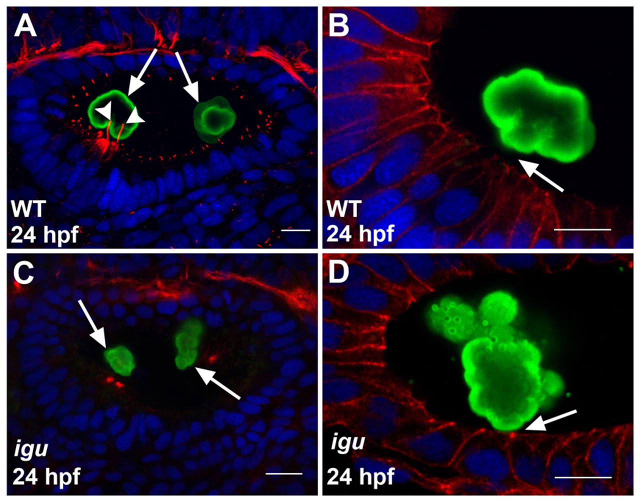
Fig. 2 Motile cilia are crucial for proper otolith formation. (A) Wild-type zebrafish ear showing the deposition of the two otoliths (green, arrows) at the poles of the otic vesicle. Note the two kinocilia (red, arrowheads) bearing the anterior otolith. (B) High-resolution image of a wild-type ear showing the anterior otolith (green). Note the gap (arrow) between the otolith surface and the apical membranes of the hair cells (red). (C) Ear of an igu mutant embryo, showing irregularly shaped otoliths (green) at the two poles (arrows) and the complete absence of cilia. (D) High-resolution image of an ear of an igu mutant showing an irregular otolith (green) attached to the apical membranes of the hair cells (red, arrow). Otoliths of embryos shown in A-D were stained with antibodies to Starmaker (Stm) (green), a component of the otoliths (Sollner et al., 2003), cilia in A and C with anti-acetylated tubulin antibodies (red), and cell membranes in B and D with anti-β-catenin antibodies (red). Nuclei were visualized with DAPI (blue). All panels show lateral views of otic vesicles with anterior to the left. Scale bars: 10 μm.