
|
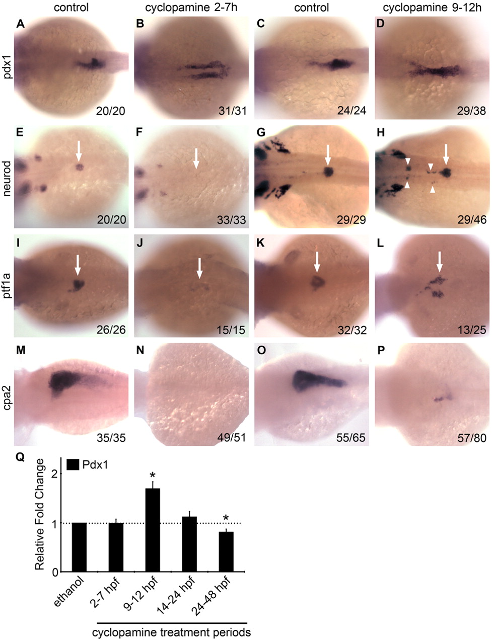
Fig. 2 Effects of transient cyclopamine treatment on exocrine and endocrine pancreas differentiation. (A-P) Dorsal views (anterior to the left) of wild-type zebrafish embryos transiently treated with cyclopamine (25 μM) either 2-7 hpf (B,F,J,N) or 9-12 hpf (D,H,L,P) versus untreated controls (A,E,I,M and C,G,K,O) and examined at 24 hpf for pdx1 (A-D) and neurod (E-H), at 48 hpf for ptf1a (I-L) and at 3 dpf for cpa2 (M-P) expression. The number of embryos displaying similar expression patterns is indicated in each case. In cyclopamine 2-7h embryos, pdx1-expressing cells fail to migrate towards the midline (A,B) and both islet and exocrine pancreas specification fail as assessed by neurod (E,F, arrows), ptf1a (I,J, arrows) and cpa2 (M,N) expression. In cyclopamine 9-12h embryos, pdx1 expression appeared expanded (C,D) and multiple clusters of neurod-expressing cells were detected at ectopic sites (G,H, arrowheads) anterior to the normal expression domain in the endogenous islet (G,H, arrow) at 24 hpf. At 48 hpf, cyclopamine 9-12h embryos show bilateral expression of ptf1a in exocrine precursors (K,L, arrows), whereas expression of cpa2 at 3 dpf in the acinar cells was reduced (O,P). (Q) Relative fold change in pdx1 expression in embryos exposed to cyclopamine transiently during various developmental periods (error bars show s.e.m.). Asterisks indicate a significant change (at least 2 s.d.) compared with control. Embryos treated during 24-48 hpf were harvested at 48 hpf and all others were harvested at 24 hpf.