
Image
|
Figure Caption
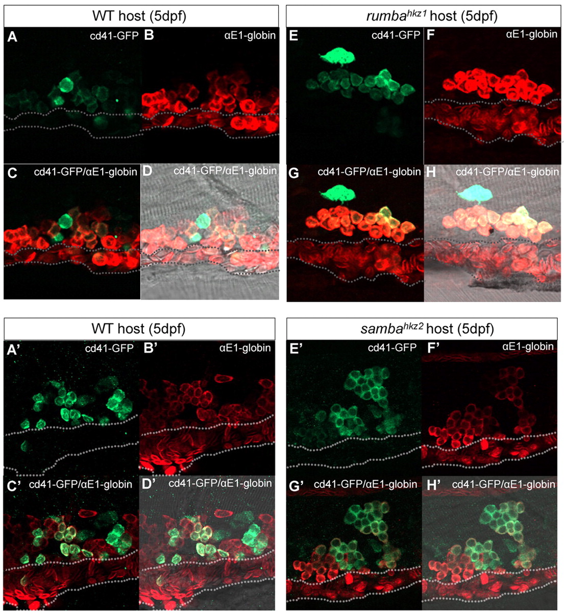
Fig. 6 Rumba and Haus3 are cell autonomously required for maintaining HSPCs in the CHT. (A-H′) Confocal images of transplantation results in wild-type (WT) zebrafish sibling hosts (A-D′), rumbahkz1-/- mutant hosts (E-H), and sambahkz2-/- mutant hosts (E′-H′). Anti-GFP staining (A,A′,E,E′) and anti-αE1-globin staining (B,B′,F,F′) in the host CHT region are merged in C, C′, G and G′, as indicated and superimposed with their respective bright field views in D, D′, H and H′. Dotted lines indicate the position of the caudal vein.
Acknowledgments
This image is the copyrighted work of the attributed author or publisher, and
ZFIN has permission only to display this image to its users.
Additional permissions should be obtained from the applicable author or publisher of the image.
Full text @ Development