
|
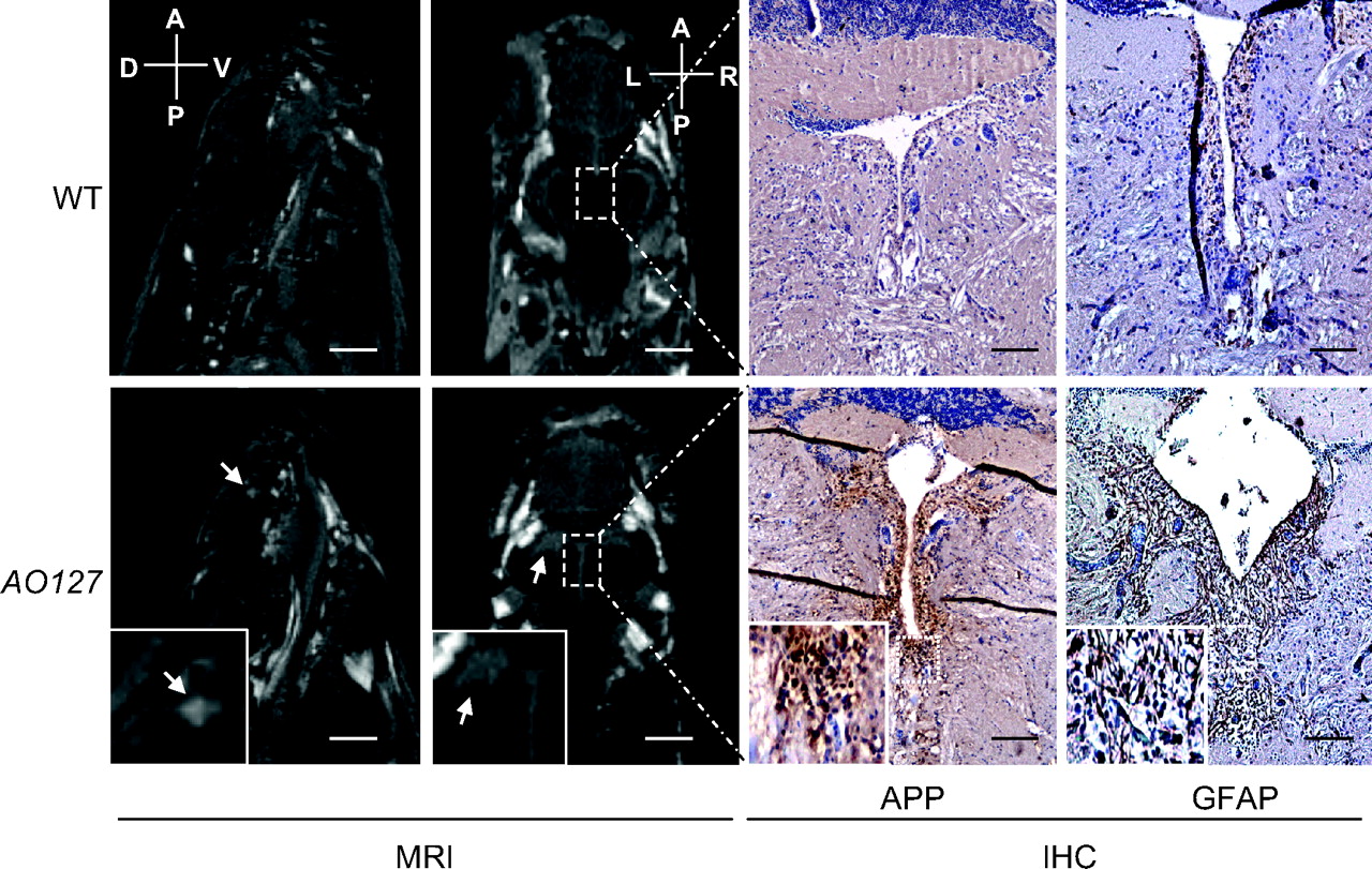
Fig. 4 White matter anomalies in AO127 mutants. T2-weighted MR images of the brain of WT and mutant AO127 adult zebrafish. Sagittal images indicate the presence of a white matter anomaly (white arrow) in telencephalon of mutant, which was not seen in WT. Coronal images show the presence of lesions adjacent to the ventricles (white arrow). IHC was carried out on the same samples used for MRI. Axonal disruptions around ventricles were seen using APP as a marker, as well as a higher number of astrocytes using GFAP as a marker, suggesting the presence of damaged neurons and inflammation around the lesions. (Scale bars: 1 mm for MR sagittal images, 500 μm for coronal images, 100 μm for immunohistochemistry.)