
Image
|
Figure Caption
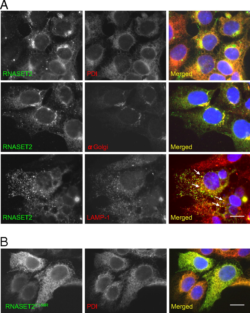
Fig. 2 Subcellular localization of RNASET2. HEK293 cells were transfected with myc-epitope tagged WT RNASET2 (A) and the mutated version, RNASET2 C184R (B). Immunofluorescence for endoplasmic reticulum (PDI), Golgi, and late endosome (LAMP-1) markers show partial colocalization with WT RNASET2, suggesting that RNASET2 enters the secretory pathway. The mutated version colocalized completely with the PDI, suggesting ER retention. (Scale bar: 10 μm.)
Acknowledgments
This image is the copyrighted work of the attributed author or publisher, and
ZFIN has permission only to display this image to its users.
Additional permissions should be obtained from the applicable author or publisher of the image.
Full text @ Proc. Natl. Acad. Sci. USA