
Image
|
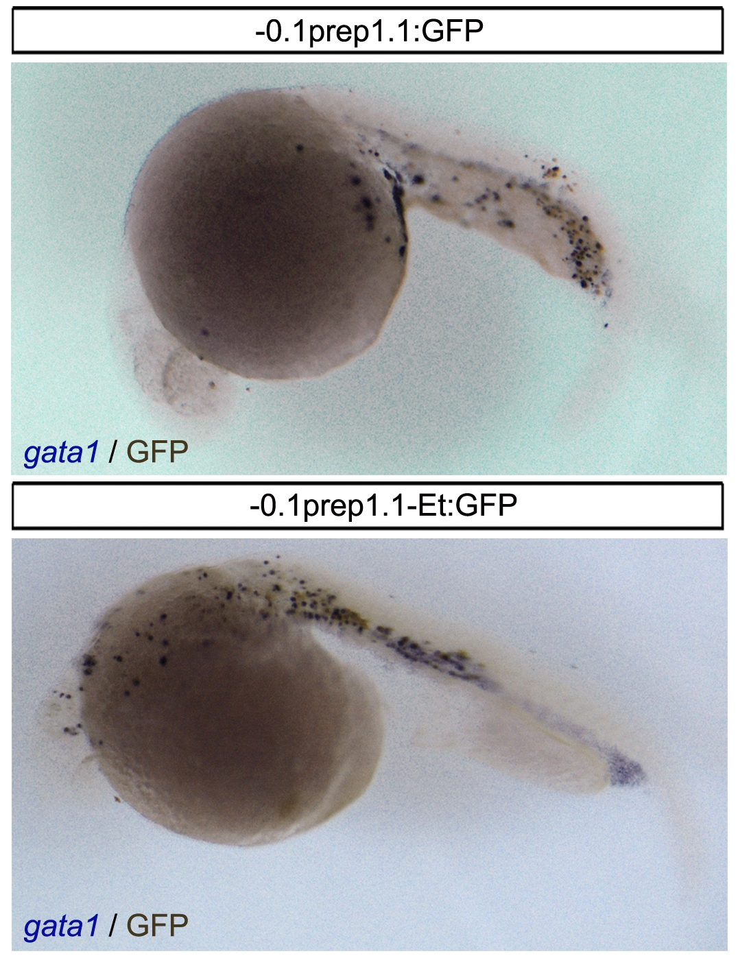
Figure Caption
Fig. 5 GFP is expressed in some gata1-positive cells.
Combined in situ hybridization for gata1 (purple) and immunohistochemistry for GFP (dark brown) in 24 hpf embryos injected with the 0.1 Kb promoter constructs (-0.1prep1.1:GFP and -0.1prep1.1-Et:GFP) reveals the co-localization of the two signals in the most of embryo.
Acknowledgments
This image is the copyrighted work of the attributed author or publisher, and
ZFIN has permission only to display this image to its users.
Additional permissions should be obtained from the applicable author or publisher of the image.
Full text @ PLoS One