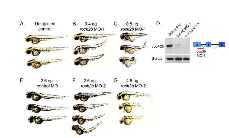
Fig. S1 Dose-dependent rock2b MO knockdown phenotypes. (A-C) Live embryos at 2 dpf. A high dose (0.8 ng) of rock2b MO-1 caused severe axis elongation defects (C) relative to a low (0.4 ng) MO dose (B) and uninjected controls (A). (D) RT-PCR was used to determine efficacy of rock2b MO-1 that targets splicing of exons 3-4 of rock2b mRNA. The 0.4 ng MO dose resulted in a partial loss of spliced transcripts, whereas the 0.8 ng dose resulted in greater depletion. β-Actin was amplified as a control. rock2b exon-intron structure and the rock2b MO-1 target site are shown in the diagram in D. (E-G) Similar dose-dependent axis elongation defects were observed in rock2b MO-2 injected embryos at 2 dpf (F,G), relative to control MO injected embryos (E).
Image
Figure Caption
Acknowledgments
This image is the copyrighted work of the attributed author or publisher, and
ZFIN has permission only to display this image to its users.
Additional permissions should be obtained from the applicable author or publisher of the image.
Full text @ Development

