
|
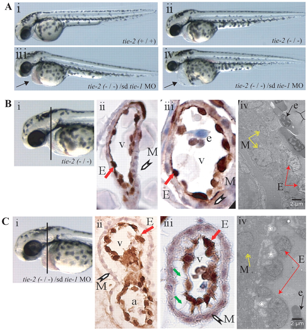
Fig. 2 Loss of tie-1 function in tie-2 mutants results in severe defects in endocardial-myocardial contact. (A) Wild type (i) and tie-2 mutants (ii) show no cardiac defects, whereas tie-1 morpholino knockdown in tie-2 mutants (iii,iv) leads to blood accumulation (arrow in iii) in the pericardial sac or to cardiac edema (arrow in iv) and arrested blood circulation (iv). (B) Immunohistochemistry using an anti-GFP antibody on heart cross-sections from 3-dpf tie-2 mutants in tg(fli1:EGFP)y1 background (i–iii) reveals no defects in myocardial (white arrowhead) and endocardial (red arrow) contact. (Biv) See below. (C) Immunohistochemistry using an anti-GFP antibody on heart cross-sections from 3-dpf tie-2 mutants in the tg(fli1:EGFP)y1 background injected with sd-tie-1 MO (i–iii) reveals defects in myocardial (white arrowhead) and endocardial (red arrow) contacts. Endothelial cells exhibit thin cytoplasmic protrusions (green arrows) attached to the myocardium. (Biv,Civ) EM studies demonstrate defects (depicted by white asterisks) in endocardial (red arrows) and myocardial (yellow arrows) lining in tie-2 mutants injected with sd-tie-1 MO (Civ) but not in control tie-2 mutants (Biv). Black arrow depicts an erythrocyte. M, myocardium; E, endocardium; v, ventricle; e, erythrocyte; a, atrium.